Movie
Movie Controller

[English] 日本語
 Yorodumi
- EMDB-18509: Amyloid-beta 40 doublet filament from the leptomeninges of indivi... -
+ Open data

- Basic information
| Entry |  | ||||||||||||
|---|---|---|---|---|---|---|---|---|---|---|---|---|---|
| Title | Amyloid-beta 40 doublet filament from the leptomeninges of individuals with Alzheimer's disease and cerebral amyloid angiopathy | ||||||||||||
 Map data Map data | |||||||||||||
 Sample Sample |
| ||||||||||||
 Keywords Keywords |  amyloid-beta / amyloid / filaments / Abeta40 / human brain / cryo-EM / Alzheimer's disease / cerebral amyloid angiopathy / PROTEIN FIBRIL amyloid-beta / amyloid / filaments / Abeta40 / human brain / cryo-EM / Alzheimer's disease / cerebral amyloid angiopathy / PROTEIN FIBRIL | ||||||||||||
| Function / homology |  Function and homology information Function and homology informationGolgi-associated vesicle / clathrin-coated pit / heparin binding / growth cone / perikaryon / early endosome / Golgi apparatus / cell surface / endoplasmic reticulum / extracellular region / nucleusSimilarity search - Function | ||||||||||||
| Biological species |  Homo sapiens (human) Homo sapiens (human) | ||||||||||||
| Method | helical reconstruction / cryo EM / Resolution: 2.7 Å | ||||||||||||
 Authors Authors | Yang Y / Murzin AS / Peak-Chew SY / Franco C / Newell KL / Ghetti B / Goedert M / Scheres SHW | ||||||||||||
| Funding support |  United Kingdom, 3 items United Kingdom, 3 items
| ||||||||||||
 Citation Citation | Journal: Acta Neuropathol Commun / Year: 2023 Title: Cryo-EM structures of Aβ40 filaments from the leptomeninges of individuals with Alzheimer's disease and cerebral amyloid angiopathy. Authors: Yang Yang / Alexey G Murzin / Sew Peak-Chew / Catarina Franco / Holly J Garringer / Kathy L Newell / Bernardino Ghetti / Michel Goedert / Sjors H W Scheres /  Abstract: We used electron cryo-microscopy (cryo-EM) to determine the structures of Aβ40 filaments from the leptomeninges of individuals with Alzheimer's disease and cerebral amyloid angiopathy. In agreement ...We used electron cryo-microscopy (cryo-EM) to determine the structures of Aβ40 filaments from the leptomeninges of individuals with Alzheimer's disease and cerebral amyloid angiopathy. In agreement with previously reported structures, which were solved to a resolution of 4.4 Å, we found three types of filaments. However, our new structures, solved to a resolution of 2.4 Å, revealed differences in the sequence assignment that redefine the fold of Aβ40 peptides and their interactions. Filaments are made of pairs of protofilaments, the ordered core of which comprises D1-G38. The different filament types comprise one, two or three protofilament pairs. In each pair, residues H14-G37 of both protofilaments adopt an extended conformation and pack against each other in an anti-parallel fashion, held together by hydrophobic interactions and hydrogen bonds between main chains and side chains. Residues D1-H13 fold back on the adjacent parts of their own chains through both polar and non-polar interactions. There are also several additional densities of unknown identity. Sarkosyl extraction and aqueous extraction gave the same structures. By cryo-EM, parenchymal deposits of Aβ42 and blood vessel deposits of Aβ40 have distinct structures, supporting the view that Alzheimer's disease and cerebral amyloid angiopathy are different Aβ proteinopathies. | ||||||||||||
| History |
|
- Structure visualization
| Supplemental images |
|---|

- Downloads & links
-EMDB archive
| Map data | emd_18509.map.gz | 42.3 MB | EMDB map data format | |
|---|---|---|---|---|
| Header (meta data) | emd-18509-v30.xmlemd-18509.xml | 14.8 KB 14.8 KB | Display Display | EMDB header |
| FSC (resolution estimation) | emd_18509_fsc.xml | 11.4 KB | Display | FSC data file |
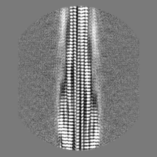
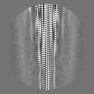
| Images | 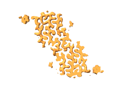 emd_18509.png emd_18509.png | 43.2 KB | ||
| Masks | emd_18509_msk_1.map | 125 MB | Mask map | |
| Filedesc metadata | emd-18509.cif.gz | 5 KB | ||
| Others | emd_18509_half_map_1.map.gzemd_18509_half_map_2.map.gz | 42.3 MB 42.3 MB | ||
| Archive directory |  http://ftp.pdbj.org/pub/emdb/structures/EMD-18509ftp://ftp.pdbj.org/pub/emdb/structures/EMD-18509 http://ftp.pdbj.org/pub/emdb/structures/EMD-18509ftp://ftp.pdbj.org/pub/emdb/structures/EMD-18509 | HTTPS FTP |
-Related structure data
| Related structure data |  8qn7MC  8qn6C M: atomic model generated by this map C: citing same article ( |
|---|---|
| Similar structure data |

-Links
| EMDB pages | EMDB (EBI/PDBe) / EMDataResource |
|---|---|
| Related items in Molecule of the Month |


-Map
| File | Download / File: emd_18509.map.gz / Format: CCP4 / Size: 125 MB / Type: IMAGE STORED AS FLOATING POINT NUMBER (4 BYTES) | ||||||||||||||||||||
|---|---|---|---|---|---|---|---|---|---|---|---|---|---|---|---|---|---|---|---|---|---|
| Voxel size | X=Y=Z: 0.744 Å | ||||||||||||||||||||
| Density |
| ||||||||||||||||||||
| Symmetry | Space group: 1 | ||||||||||||||||||||
| Details | EMDB XML:
|
-Supplemental data
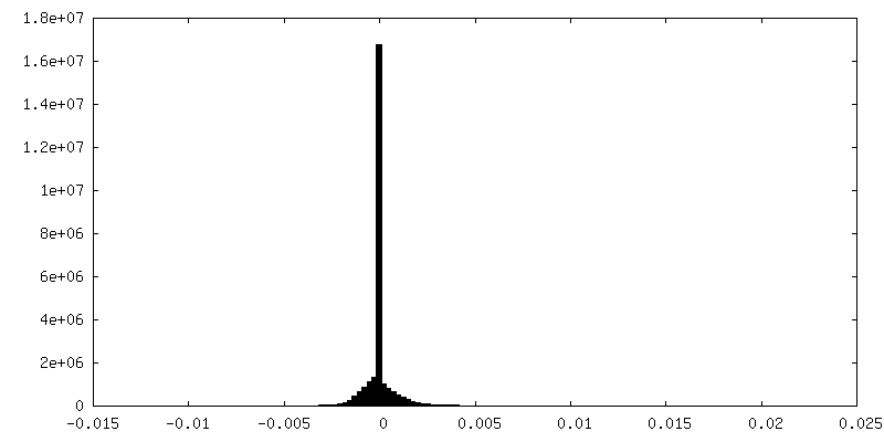
-Mask #1
| File | emd_18509_msk_1.map | ||||||||||||
|---|---|---|---|---|---|---|---|---|---|---|---|---|---|
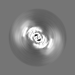
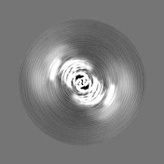
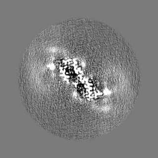
| Projections & Slices |
| ||||||||||||
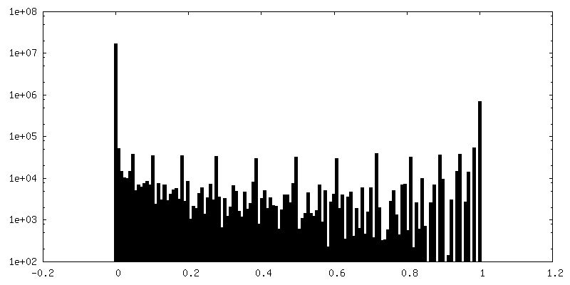
| Density Histograms |
 Z
Z Y
Y X
X


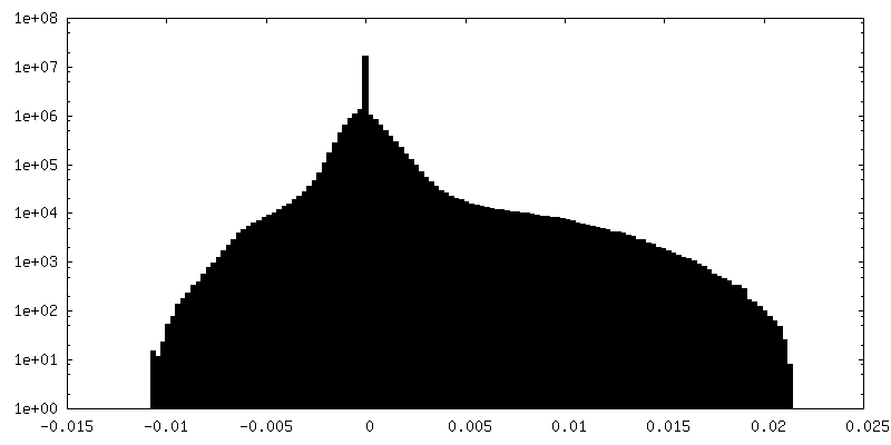
-Half map: #1
| File | emd_18509_half_map_1.map | ||||||||||||
|---|---|---|---|---|---|---|---|---|---|---|---|---|---|
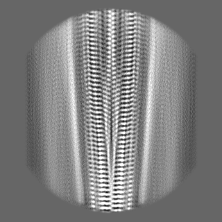
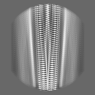
| Projections & Slices |
| ||||||||||||
| Density Histograms |


-Half map: #2
| File | emd_18509_half_map_2.map | ||||||||||||
|---|---|---|---|---|---|---|---|---|---|---|---|---|---|
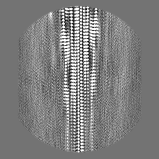
| Projections & Slices |
| ||||||||||||
| Density Histograms |


- Sample components
-Entire : Amyloid-beta 40 doublet filament extracted from the human brain w...
| Entire | Name: Amyloid-beta 40 doublet filament extracted from the human brain with Alzheimer's disease and cerebral amyloid angiopathy |
|---|---|
| Components |
|
-Supramolecule #1: Amyloid-beta 40 doublet filament extracted from the human brain w...
| Supramolecule | Name: Amyloid-beta 40 doublet filament extracted from the human brain with Alzheimer's disease and cerebral amyloid angiopathy type: tissue / ID: 1 / Parent: 0 / Macromolecule list: all |
|---|---|
| Source (natural) | Organism: Homo sapiens (human) |
-Macromolecule #1: Amyloid-beta A4 protein
| Macromolecule | Name: Amyloid-beta A4 protein / type: protein_or_peptide / ID: 1 / Number of copies: 1 / Enantiomer: LEVO |
|---|---|
| Source (natural) | Organism: Homo sapiens (human) |
| Molecular weight | Theoretical: 4.335852 KDa |
| Sequence | String: DAEFRHDSGY EVHHQKLVFF AEDVGSNKGA IIGLMVGGVV UniProtKB: Amyloid-beta A4 protein |
-Experimental details
-Structure determination
| Method | cryo EM |
|---|---|
 Processing Processing | helical reconstruction |
| Aggregation state | filament |
-Sample preparation
| Buffer | pH: 7.5 |
|---|---|
| Vitrification | Cryogen name: ETHANE |
- Electron microscopy
| Microscope | FEI TITAN KRIOS |
|---|---|
| Electron beam | Acceleration voltage: 300 kV / Electron source: FIELD EMISSION GUN |
| Electron optics | Illumination mode: FLOOD BEAM / Imaging mode: BRIGHT FIELDBright-field microscopy / Nominal defocus max: 2.4 µm / Nominal defocus min: 1.0 µm |
| Image recording | Film or detector model: FEI FALCON IV (4k x 4k) / Average electron dose: 40.0 e/Å2 |
| Experimental equipment |  Model: Titan Krios / Image courtesy: FEI Company |
-Image processing
| Startup model | Type of model: PDB ENTRY PDB model - PDB ID: |
|---|---|
| Final angle assignment | Type: NOT APPLICABLE / Software - Name: RELION |
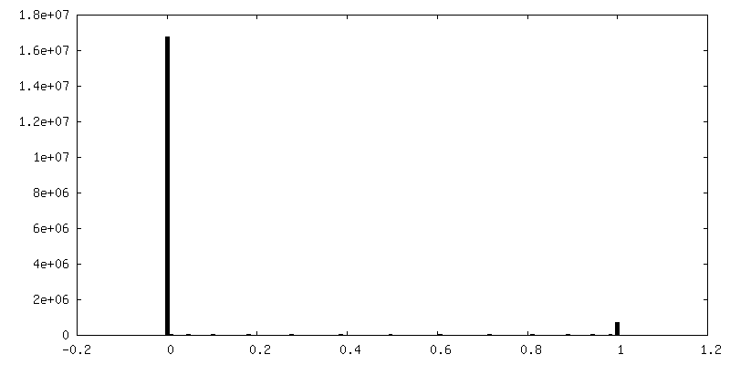
| Final reconstruction | Applied symmetry - Helical parameters - Δz: 2.44 Å Applied symmetry - Helical parameters - Δ&Phi: -179.53 ° Applied symmetry - Helical parameters - Axial symmetry: C1 (asymmetric) Resolution.type: BY AUTHOR / Resolution: 2.7 Å / Resolution method: FSC 0.143 CUT-OFF / Software - Name: RELION / Number images used: 32218 |
| FSC plot (resolution estimation) | 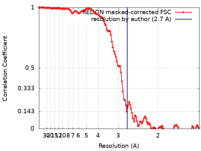 |