 Movie
Movie Controller


+ Open data

- Basic information
| Entry | 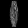 | ||||||||||||||||||
|---|---|---|---|---|---|---|---|---|---|---|---|---|---|---|---|---|---|---|---|
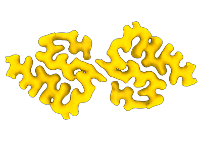
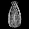
| Title | Murine type II Abeta fibril from APP23 mouse | ||||||||||||||||||
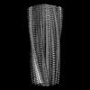
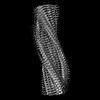
 Map data Map data | Ex vivo Abeta42 fibril from APP23 mouse brain. | ||||||||||||||||||
 Sample Sample |
| ||||||||||||||||||
 Keywords Keywords | Amyloid fibril / PROTEIN FIBRIL | ||||||||||||||||||
| Function / homology |  Function and homology information Function and homology informationregulation of epidermal growth factor-activated receptor activity / cytosolic mRNA polyadenylation / collateral sprouting in absence of injury / microglia development / regulation of synapse structure or activity / regulation of Wnt signaling pathway / Formyl peptide receptors bind formyl peptides and many other ligands / axo-dendritic transport / synaptic assembly at neuromuscular junction / signaling receptor activator activity ...regulation of epidermal growth factor-activated receptor activity / cytosolic mRNA polyadenylation / collateral sprouting in absence of injury / microglia development / regulation of synapse structure or activity / regulation of Wnt signaling pathway / Formyl peptide receptors bind formyl peptides and many other ligands / axo-dendritic transport / synaptic assembly at neuromuscular junction / signaling receptor activator activity / smooth endoplasmic reticulum calcium ion homeostasis / axon midline choice point recognition / astrocyte activation involved in immune response / regulation of spontaneous synaptic transmission / mating behavior / NMDA selective glutamate receptor signaling pathway / ciliary rootlet / Lysosome Vesicle Biogenesis / PTB domain binding / Golgi-associated vesicle / positive regulation of amyloid fibril formation / neuron remodeling / : / Insertion of tail-anchored proteins into the endoplasmic reticulum membrane / Deregulated CDK5 triggers multiple neurodegenerative pathways in Alzheimer's disease models / suckling behavior / nuclear envelope lumen / dendrite development / COPII-coated ER to Golgi transport vesicle / presynaptic active zone / modulation of excitatory postsynaptic potential / TRAF6 mediated NF-kB activation / Advanced glycosylation endproduct receptor signaling / neuromuscular process controlling balance / The NLRP3 inflammasome / regulation of presynapse assembly / transition metal ion binding / negative regulation of long-term synaptic potentiation / regulation of multicellular organism growth / intracellular copper ion homeostasis / negative regulation of neuron differentiation / ECM proteoglycans / smooth endoplasmic reticulum / positive regulation of T cell migration / spindle midzone / Purinergic signaling in leishmaniasis infection / positive regulation of calcium-mediated signaling / protein serine/threonine kinase binding / positive regulation of chemokine production / clathrin-coated pit / regulation of peptidyl-tyrosine phosphorylation / forebrain development / Notch signaling pathway / Mitochondrial protein degradation / neuron projection maintenance / positive regulation of G2/M transition of mitotic cell cycle / positive regulation of protein metabolic process / ionotropic glutamate receptor signaling pathway / positive regulation of glycolytic process / cholesterol metabolic process / positive regulation of mitotic cell cycle / response to interleukin-1 / adult locomotory behavior / extracellular matrix organization / axonogenesis / platelet alpha granule lumen / trans-Golgi network membrane / positive regulation of peptidyl-threonine phosphorylation / dendritic shaft / learning / positive regulation of interleukin-1 beta production / positive regulation of long-term synaptic potentiation / locomotory behavior / central nervous system development / endosome lumen / astrocyte activation / positive regulation of JNK cascade / Post-translational protein phosphorylation / synapse organization / regulation of long-term neuronal synaptic plasticity / microglial cell activation / TAK1-dependent IKK and NF-kappa-B activation / visual learning / serine-type endopeptidase inhibitor activity / neuromuscular junction / recycling endosome / cognition / neuron cellular homeostasis / Golgi lumen / positive regulation of inflammatory response / positive regulation of non-canonical NF-kappaB signal transduction / endocytosis / cellular response to amyloid-beta / G2/M transition of mitotic cell cycle / Regulation of Insulin-like Growth Factor (IGF) transport and uptake by Insulin-like Growth Factor Binding Proteins (IGFBPs) / positive regulation of interleukin-6 production / positive regulation of tumor necrosis factor production / neuron projection development / cell-cell junction / synaptic vesicle Similarity search - Function | ||||||||||||||||||
| Biological species |   Homo sapiens (human) Homo sapiens (human) | ||||||||||||||||||
| Method | helical reconstruction / cryo EM / Resolution: 3.0 Å | ||||||||||||||||||
 Authors Authors | Zielinski M / Peralta Reyes FS / Gremer L / Schemmert S / Frieg B / Willuweit A / Donner L / Elvers M / Nilsson LNG / Syvanen S ...Zielinski M / Peralta Reyes FS / Gremer L / Schemmert S / Frieg B / Willuweit A / Donner L / Elvers M / Nilsson LNG / Syvanen S / Sehlin D / Ingelsson M / Willbold D / Schroeder GF | ||||||||||||||||||
| Funding support |  Germany, Germany,  Sweden, 5 items Sweden, 5 items
| ||||||||||||||||||
 Citation Citation | Journal: Nat Neurosci / Year: 2023 Title: Cryo-EM of Aβ fibrils from mouse models find tg-APP fibrils resemble those found in patients with sporadic Alzheimer's disease. Authors: Mara Zielinski / Fernanda S Peralta Reyes / Lothar Gremer / Sarah Schemmert / Benedikt Frieg / Luisa U Schäfer / Antje Willuweit / Lili Donner / Margitta Elvers / Lars N G Nilsson / Stina ...Authors: Mara Zielinski / Fernanda S Peralta Reyes / Lothar Gremer / Sarah Schemmert / Benedikt Frieg / Luisa U Schäfer / Antje Willuweit / Lili Donner / Margitta Elvers / Lars N G Nilsson / Stina Syvänen / Dag Sehlin / Martin Ingelsson / Dieter Willbold / Gunnar F Schröder /   Abstract: The use of transgenic mice displaying amyloid-β (Aβ) brain pathology has been essential for the preclinical assessment of new treatment strategies for Alzheimer's disease. However, the properties ...The use of transgenic mice displaying amyloid-β (Aβ) brain pathology has been essential for the preclinical assessment of new treatment strategies for Alzheimer's disease. However, the properties of Aβ in such mice have not been systematically compared to Aβ in the brains of patients with Alzheimer's disease. Here, we determined the structures of nine ex vivo Aβ fibrils from six different mouse models by cryogenic-electron microscopy. We found novel Aβ fibril structures in the APP/PS1, ARTE10 and tg-SwDI models, whereas the human type II filament fold was found in the ARTE10, tg-APP and APP23 models. The tg-APP mice showed an Aβ fibril whose structure resembles the human type I filament found in patients with sporadic Alzheimer's disease. A detailed assessment of the Aβ fibril structure is key to the selection of adequate mouse models for the preclinical development of novel plaque-targeting therapeutics and positron emission tomography imaging tracers in Alzheimer's disease. | ||||||||||||||||||
| History |
|
- Structure visualization
| Supplemental images |
|---|
- Downloads & links
-EMDB archive
| Map data | emd_16942.map.gz | 10.4 MB | EMDB map data format | |
|---|---|---|---|---|
| Header (meta data) | emd-16942-v30.xmlemd-16942.xml | 15.7 KB 15.7 KB | Display Display | EMDB header |
| FSC (resolution estimation) | emd_16942_fsc.xml | 10.7 KB | Display | FSC data file |
| Images |  emd_16942.png emd_16942.png | 59 KB | ||
| Filedesc metadata | emd-16942.cif.gz | 5.3 KB | ||
| Others | emd_16942_half_map_1.map.gzemd_16942_half_map_2.map.gz | 78.9 MB 78.8 MB | ||
| Archive directory |  http://ftp.pdbj.org/pub/emdb/structures/EMD-16942ftp://ftp.pdbj.org/pub/emdb/structures/EMD-16942 http://ftp.pdbj.org/pub/emdb/structures/EMD-16942ftp://ftp.pdbj.org/pub/emdb/structures/EMD-16942 | HTTPS FTP |
-Validation report
| Summary document | emd_16942_validation.pdf.gz | 697.4 KB | Display | EMDB validaton report |
|---|---|---|---|---|
| Full document | emd_16942_full_validation.pdf.gz | 697 KB | Display | |
| Data in XML | emd_16942_validation.xml.gz | 18.2 KB | Display | |
| Data in CIF | emd_16942_validation.cif.gz | 23.7 KB | Display | |
| Arichive directory | https://ftp.pdbj.org/pub/emdb/validation_reports/EMD-16942ftp://ftp.pdbj.org/pub/emdb/validation_reports/EMD-16942 | HTTPS FTP |
-Related structure data
| Related structure data | 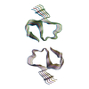 8ol2MC 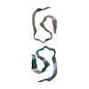 8ol3C 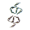 8ol5C 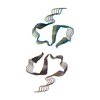 8ol6C 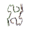 8ol7C 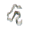 8olgC  8olnC  8oloC  8olqC M: atomic model generated by this map C: citing same article ( |
|---|---|
| Similar structure data |
-Links
| EMDB pages | EMDB (EBI/PDBe) / EMDataResource |
|---|---|
| Related items in Molecule of the Month |


-Map
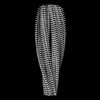
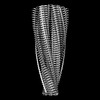
| File | Download / File: emd_16942.map.gz / Format: CCP4 / Size: 103 MB / Type: IMAGE STORED AS FLOATING POINT NUMBER (4 BYTES) | ||||||||||||||||||||||||||||||||||||
|---|---|---|---|---|---|---|---|---|---|---|---|---|---|---|---|---|---|---|---|---|---|---|---|---|---|---|---|---|---|---|---|---|---|---|---|---|---|
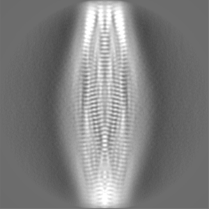
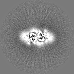
| Annotation | Ex vivo Abeta42 fibril from APP23 mouse brain. | ||||||||||||||||||||||||||||||||||||
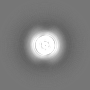
| Projections & slices | Image control
Images are generated by Spider. | ||||||||||||||||||||||||||||||||||||
| Voxel size | X=Y=Z: 0.808 Å | ||||||||||||||||||||||||||||||||||||
| Density |
| ||||||||||||||||||||||||||||||||||||
| Symmetry | Space group: 1 | ||||||||||||||||||||||||||||||||||||
| Details | EMDB XML:
|
 Z (Sec.)
 Y (Row.)
 X (Col.)


-Supplemental data
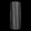
-Half map: Ex vivo Abeta42 fibril from APP23 mouse brain. Half map 2.
| File | emd_16942_half_map_1.map | ||||||||||||
|---|---|---|---|---|---|---|---|---|---|---|---|---|---|
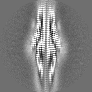
| Annotation | Ex vivo Abeta42 fibril from APP23 mouse brain. Half map 2. | ||||||||||||
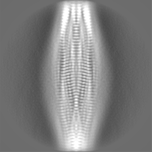
| Projections & Slices |
| ||||||||||||
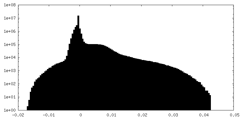
| Density Histograms |


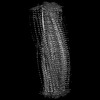
-Half map: Ex vivo Abeta42 fibril from APP23 mouse brain. Half map 1.
| File | emd_16942_half_map_2.map | ||||||||||||
|---|---|---|---|---|---|---|---|---|---|---|---|---|---|
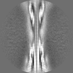
| Annotation | Ex vivo Abeta42 fibril from APP23 mouse brain. Half map 1. | ||||||||||||
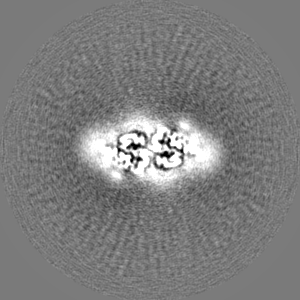
| Projections & Slices |
| ||||||||||||
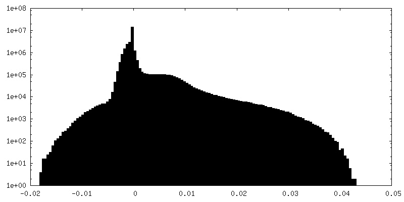
| Density Histograms |


- Sample components
-Entire : Amyloid fibril of amyloid-beta
| Entire | Name: Amyloid fibril of amyloid-beta |
|---|---|
| Components |
|
-Supramolecule #1: Amyloid fibril of amyloid-beta
| Supramolecule | Name: Amyloid fibril of amyloid-beta / type: complex / ID: 1 / Parent: 0 / Macromolecule list: all |
|---|---|
| Source (natural) | Organism: |
| Molecular weight | Theoretical: 20 kDa/nm |
-Macromolecule #1: Amyloid-beta protein 42
| Macromolecule | Name: Amyloid-beta protein 42 / type: protein_or_peptide / ID: 1 / Number of copies: 10 / Enantiomer: LEVO |
|---|---|
| Source (natural) | Organism: Homo sapiens (human) / Organ: Brain |
| Molecular weight | Theoretical: 4.520087 KDa |
| Recombinant expression | Organism: |
| Sequence | String: DAEFRHDSGY EVHHQKLVFF AEDVGSNKGA IIGLMVGGVV IA UniProtKB: Amyloid-beta precursor protein |
-Experimental details
-Structure determination
| Method | cryo EM |
|---|---|
 Processing Processing | helical reconstruction |
| Aggregation state | filament |
-Sample preparation
| Buffer | pH: 7.4 |
|---|---|
| Vitrification | Cryogen name: ETHANE / Instrument: FEI VITROBOT MARK IV |
- Electron microscopy
| Microscope | TFS KRIOS |
|---|---|
| Image recording | Film or detector model: FEI FALCON IV (4k x 4k) / Number grids imaged: 1 / Number real images: 11622 / Average electron dose: 40.0 e/Å2 |
| Electron beam | Acceleration voltage: 300 kV / Electron source:  FIELD EMISSION GUN FIELD EMISSION GUN |
| Electron optics | Illumination mode: FLOOD BEAM / Imaging mode: BRIGHT FIELD / Nominal defocus max: 3.0 µm / Nominal defocus min: 0.5 µm |
| Experimental equipment |  Model: Titan Krios / Image courtesy: FEI Company |
-Image processing
| Final reconstruction | Applied symmetry - Helical parameters - Δz: 4.77 Å Applied symmetry - Helical parameters - Δ&Phi: -3.23 ° Applied symmetry - Helical parameters - Axial symmetry: C2 (2 fold cyclic) Algorithm: FOURIER SPACE / Resolution.type: BY AUTHOR / Resolution: 3.0 Å / Resolution method: FSC 0.143 CUT-OFF / Software - Name: RELION (ver. 3.1) / Number images used: 119710 |
|---|---|
| Startup model | Type of model: OTHER / Details: cylinder. |
| Final angle assignment | Type: NOT APPLICABLE |
| FSC plot (resolution estimation) | 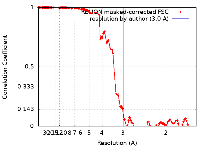 |
