 Movie
Movie Controller

[English] 日本語
 Yorodumi
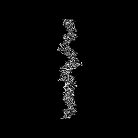
- EMDB-16683: Cryo-EM structure of the CupE pilus from Pseudomonas aeruginosa -
+ Open data

- Basic information
| Entry | 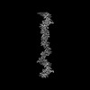 | ||||||||||||||||||||||||
|---|---|---|---|---|---|---|---|---|---|---|---|---|---|---|---|---|---|---|---|---|---|---|---|---|---|
| Title | Cryo-EM structure of the CupE pilus from Pseudomonas aeruginosa | ||||||||||||||||||||||||
 Map data Map data | |||||||||||||||||||||||||
 Sample Sample |
| ||||||||||||||||||||||||
 Keywords Keywords | Pseudomonas aeruginosa / pilus / Chaperone-Usher / Chaperone-Usher Pathway / adhesin / biofilm / PROTEIN FIBRIL | ||||||||||||||||||||||||
| Function / homology | : / Spore coat protein U / Spore Coat Protein U domain / Spore Coat Protein U domain / Spore coat protein U/FanG domain-containing protein Function and homology information Function and homology information | ||||||||||||||||||||||||
| Biological species |   Pseudomonas aeruginosa (bacteria) / Pseudomonas aeruginosa PAO1 (bacteria) Pseudomonas aeruginosa (bacteria) / Pseudomonas aeruginosa PAO1 (bacteria) | ||||||||||||||||||||||||
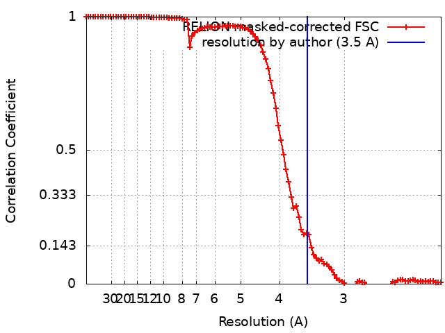
| Method | helical reconstruction / cryo EM / Resolution: 3.5 Å | ||||||||||||||||||||||||
 Authors Authors | Boehning J / Bharat TAM | ||||||||||||||||||||||||
| Funding support |  United Kingdom, United Kingdom,  United States, 7 items United States, 7 items
| ||||||||||||||||||||||||
 Citation Citation | Journal: PLoS Pathog / Year: 2023 Title: Architecture of the biofilm-associated archaic Chaperone-Usher pilus CupE from Pseudomonas aeruginosa. Authors: Jan Böhning / Adrian W Dobbelstein / Nina Sulkowski / Kira Eilers / Andriko von Kügelgen / Abul K Tarafder / Sew-Yeu Peak-Chew / Mark Skehel / Vikram Alva / Alain Filloux / Tanmay A M Bharat /  Abstract: Chaperone-Usher Pathway (CUP) pili are major adhesins in Gram-negative bacteria, mediating bacterial adherence to biotic and abiotic surfaces. While classical CUP pili have been extensively ...Chaperone-Usher Pathway (CUP) pili are major adhesins in Gram-negative bacteria, mediating bacterial adherence to biotic and abiotic surfaces. While classical CUP pili have been extensively characterized, little is known about so-called archaic CUP pili, which are phylogenetically widespread and promote biofilm formation by several human pathogens. In this study, we present the electron cryomicroscopy structure of the archaic CupE pilus from the opportunistic human pathogen Pseudomonas aeruginosa. We show that CupE1 subunits within the pilus are arranged in a zigzag architecture, containing an N-terminal donor β-strand extending from each subunit into the next, where it is anchored by hydrophobic interactions, with comparatively weaker interactions at the rest of the inter-subunit interface. Imaging CupE pili on the surface of P. aeruginosa cells using electron cryotomography shows that CupE pili adopt variable curvatures in response to their environment, which might facilitate their role in promoting cellular attachment. Finally, bioinformatic analysis shows the widespread abundance of cupE genes in isolates of P. aeruginosa and the co-occurrence of cupE with other cup clusters, suggesting interdependence of cup pili in regulating bacterial adherence within biofilms. Taken together, our study provides insights into the architecture of archaic CUP pili, providing a structural basis for understanding their role in promoting cellular adhesion and biofilm formation in P. aeruginosa. | ||||||||||||||||||||||||
| History |
|
- Structure visualization
| Supplemental images |
|---|

- Downloads & links
-EMDB archive
| Map data | emd_16683.map.gz | 5.1 MB | EMDB map data format | |
|---|---|---|---|---|
| Header (meta data) | emd-16683-v30.xmlemd-16683.xml | 17.1 KB 17.1 KB | Display Display | EMDB header |
| FSC (resolution estimation) | emd_16683_fsc.xml | 10 KB | Display | FSC data file |
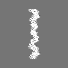
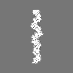
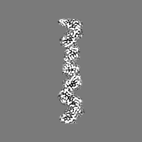
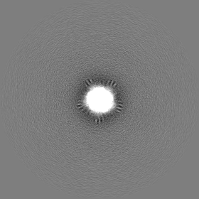
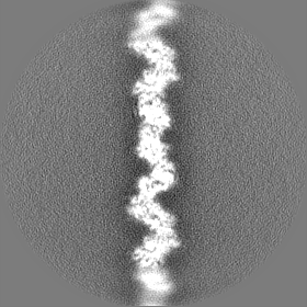
| Images | 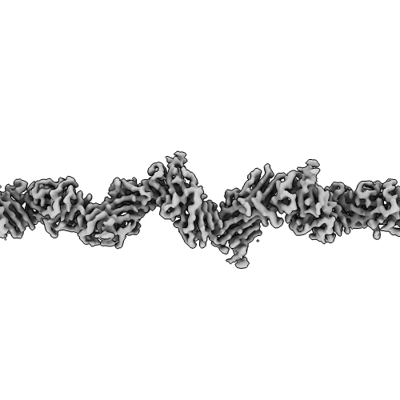 emd_16683.png emd_16683.png | 46.2 KB | ||
| Filedesc metadata | emd-16683.cif.gz | 5.6 KB | ||
| Others | emd_16683_half_map_1.map.gzemd_16683_half_map_2.map.gz | 65.6 MB 65.5 MB | ||
| Archive directory |  http://ftp.pdbj.org/pub/emdb/structures/EMD-16683ftp://ftp.pdbj.org/pub/emdb/structures/EMD-16683 http://ftp.pdbj.org/pub/emdb/structures/EMD-16683ftp://ftp.pdbj.org/pub/emdb/structures/EMD-16683 | HTTPS FTP |
-Related structure data
| Related structure data |  8cioMC M: atomic model generated by this map C: citing same article ( |
|---|---|
| Similar structure data |
-Links
| EMDB pages | EMDB (EBI/PDBe) / EMDataResource |
|---|
-Map
| File | Download / File: emd_16683.map.gz / Format: CCP4 / Size: 83.7 MB / Type: IMAGE STORED AS FLOATING POINT NUMBER (4 BYTES) | ||||||||||||||||||||||||||||||||||||
|---|---|---|---|---|---|---|---|---|---|---|---|---|---|---|---|---|---|---|---|---|---|---|---|---|---|---|---|---|---|---|---|---|---|---|---|---|---|
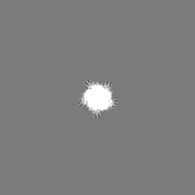
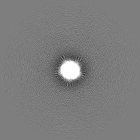
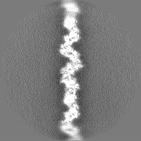
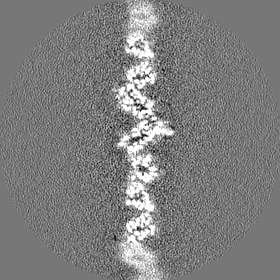
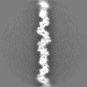
| Projections & slices | Image control
Images are generated by Spider. | ||||||||||||||||||||||||||||||||||||
| Voxel size | X=Y=Z: 1.092 Å | ||||||||||||||||||||||||||||||||||||
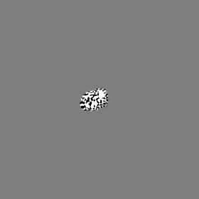
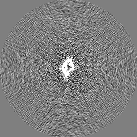
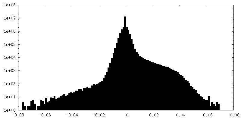
| Density |
| ||||||||||||||||||||||||||||||||||||
| Symmetry | Space group: 1 | ||||||||||||||||||||||||||||||||||||
| Details | EMDB XML:
|
 Z (Sec.)
 Y (Row.)
 X (Col.)


-Supplemental data
-Half map: #1
| File | emd_16683_half_map_1.map | ||||||||||||
|---|---|---|---|---|---|---|---|---|---|---|---|---|---|
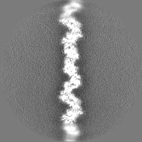
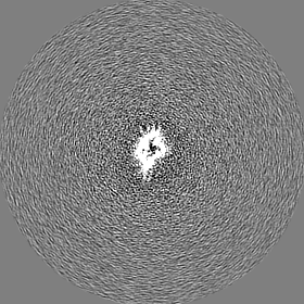
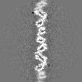
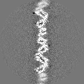
| Projections & Slices |
| ||||||||||||
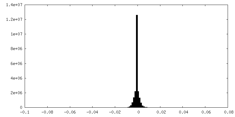
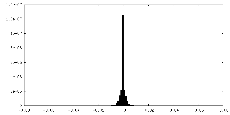
| Density Histograms |
-Half map: #2
| File | emd_16683_half_map_2.map | ||||||||||||
|---|---|---|---|---|---|---|---|---|---|---|---|---|---|
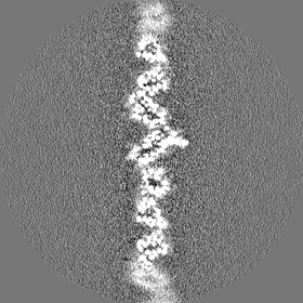
| Projections & Slices |
| ||||||||||||
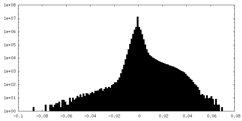
| Density Histograms |
- Sample components
-Entire : CupE pilus
| Entire | Name: CupE pilus |
|---|---|
| Components |
|
-Supramolecule #1: CupE pilus
| Supramolecule | Name: CupE pilus / type: complex / ID: 1 / Parent: 0 / Macromolecule list: all |
|---|---|
| Source (natural) | Organism: Pseudomonas aeruginosa (bacteria) / Strain: PAO1 mutant / Location in cell: Cell surface |
-Macromolecule #1: SCPU domain-containing protein
| Macromolecule | Name: SCPU domain-containing protein / type: protein_or_peptide / ID: 1 / Number of copies: 5 / Enantiomer: LEVO |
|---|---|
| Source (natural) | Organism: Pseudomonas aeruginosa PAO1 (bacteria) |
| Molecular weight | Theoretical: 16.127815 KDa |
| Sequence | String: AGTLIGQVGV QMVIGAGCTI INGSVSGGIN QWGTLDFGSH SDLTNVVDAQ TVGTSGNIQI QCSTGLTPSL TVNAGLHASG GQRYMQNTT TTSSTIAYNI YSDAARSALI QANTPVDISS VSTGTAVNIP LYGRVVPTGQ STPTPTAGTY TDTLLVTIAW UniProtKB: Spore coat protein U/FanG domain-containing protein |
-Experimental details
-Structure determination
| Method | cryo EM |
|---|---|
 Processing Processing | helical reconstruction |
| Aggregation state | filament |
-Sample preparation
| Buffer | pH: 7.4 / Details: Phosphate-buffered saline |
|---|---|
| Grid | Model: Quantifoil R2/2 / Material: COPPER/RHODIUM / Mesh: 200 / Support film - Material: CARBON / Support film - topology: HOLEY ARRAY / Pretreatment - Type: GLOW DISCHARGE / Pretreatment - Time: 20 sec. / Details: 15 mA |
| Vitrification | Cryogen name: ETHANE / Chamber humidity: 100 % / Chamber temperature: 283 K / Instrument: FEI VITROBOT MARK IV |
- Electron microscopy
| Microscope | FEI TITAN KRIOS |
|---|---|
| Specialist optics | Energy filter - Name: GIF Bioquantum / Energy filter - Slit width: 20 eV |
| Image recording | Film or detector model: GATAN K3 (6k x 4k) / Number grids imaged: 1 / Average electron dose: 46.0 e/Å2 |
| Electron beam | Acceleration voltage: 300 kV / Electron source:  FIELD EMISSION GUN FIELD EMISSION GUN |
| Electron optics | Illumination mode: FLOOD BEAM / Imaging mode: BRIGHT FIELD / Cs: 2.7 mm / Nominal defocus max: 2.5 µm / Nominal defocus min: 1.0 µm / Nominal magnification: 81000 |
| Sample stage | Specimen holder model: FEI TITAN KRIOS AUTOGRID HOLDER / Cooling holder cryogen: NITROGEN |
| Experimental equipment |  Model: Titan Krios / Image courtesy: FEI Company |
+Image processing
-Atomic model buiding 1
| Details | Initial local fitting of a homology model was done using ChimeraX, and the structure manually re-built in Coot. |
|---|---|
| Refinement | Space: REAL / Protocol: AB INITIO MODEL |
| Output model | PDB-8cio: |
