 Movie
Movie Controller

[English] 日本語
 Yorodumi
- EMDB-16520: Omadacycline and spectinomycin bound to the 30S ribosomal subunit head -
+ Open data

- Basic information
| Entry |  | ||||||||||||
|---|---|---|---|---|---|---|---|---|---|---|---|---|---|
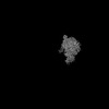
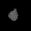
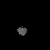
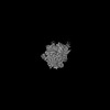
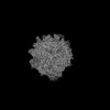
| Title | Omadacycline and spectinomycin bound to the 30S ribosomal subunit head | ||||||||||||
 Map data Map data | |||||||||||||
 Sample Sample |
| ||||||||||||
 Keywords Keywords | Ribosome / antibiotics / spectinomycin / omadacycline | ||||||||||||
| Function / homology |  Function and homology information Function and homology informationtranscription antitermination factor activity, RNA binding / regulation of DNA-templated transcription elongation / transcription elongation factor complex / transcription antitermination / maintenance of translational fidelity / ribosomal small subunit assembly / ribosome biogenesis / small ribosomal subunit / cytosolic small ribosomal subunit / cytoplasmic translation ...transcription antitermination factor activity, RNA binding / regulation of DNA-templated transcription elongation / transcription elongation factor complex / transcription antitermination / maintenance of translational fidelity / ribosomal small subunit assembly / ribosome biogenesis / small ribosomal subunit / cytosolic small ribosomal subunit / cytoplasmic translation / tRNA binding / negative regulation of translation / rRNA binding / structural constituent of ribosome / ribosome / translation / response to antibiotic / mRNA binding / RNA binding / membrane / cytoplasm / cytosol Similarity search - Function | ||||||||||||
| Biological species |  | ||||||||||||
| Method | single particle reconstruction / cryo EM / Resolution: 2.06 Å | ||||||||||||
 Authors Authors | Paternoga H / Crowe-McAuliffe C / Wilson DN | ||||||||||||
| Funding support | European Union, 3 items
| ||||||||||||
 Citation Citation | Journal: Nat Struct Mol Biol / Year: 2023 Title: Structural conservation of antibiotic interaction with ribosomes. Authors: Helge Paternoga / Caillan Crowe-McAuliffe / Lars V Bock / Timm O Koller / Martino Morici / Bertrand Beckert / Alexander G Myasnikov / Helmut Grubmüller / Jiří Nováček / Daniel N Wilson /    Abstract: The ribosome is a major target for clinically used antibiotics, but multidrug resistant pathogenic bacteria are making our current arsenal of antimicrobials obsolete. Here we present cryo-electron- ...The ribosome is a major target for clinically used antibiotics, but multidrug resistant pathogenic bacteria are making our current arsenal of antimicrobials obsolete. Here we present cryo-electron-microscopy structures of 17 distinct compounds from six different antibiotic classes bound to the bacterial ribosome at resolutions ranging from 1.6 to 2.2 Å. The improved resolution enables a precise description of antibiotic-ribosome interactions, encompassing solvent networks that mediate multiple additional interactions between the drugs and their target. Our results reveal a high structural conservation in the binding mode between antibiotics with the same scaffold, including ordered water molecules. Water molecules are visualized within the antibiotic binding sites that are preordered, become ordered in the presence of the drug and that are physically displaced on drug binding. Insight into RNA-ligand interactions will facilitate development of new antimicrobial agents, as well as other RNA-targeting therapies. | ||||||||||||
| History |
|
- Structure visualization
| Supplemental images |
|---|

- Downloads & links
-EMDB archive
| Map data | emd_16520.map.gz | 26.6 MB | EMDB map data format | |
|---|---|---|---|---|
| Header (meta data) | emd-16520-v30.xmlemd-16520.xml | 33.4 KB 33.4 KB | Display Display | EMDB header |
| Images | 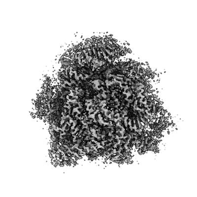 emd_16520.png emd_16520.png | 118.5 KB | ||
| Others | emd_16520_additional_1.map.gzemd_16520_half_map_1.map.gzemd_16520_half_map_2.map.gz | 402.5 MB 408.7 MB 408.7 MB | ||
| Archive directory |  http://ftp.pdbj.org/pub/emdb/structures/EMD-16520ftp://ftp.pdbj.org/pub/emdb/structures/EMD-16520 http://ftp.pdbj.org/pub/emdb/structures/EMD-16520ftp://ftp.pdbj.org/pub/emdb/structures/EMD-16520 | HTTPS FTP |
-Related structure data
| Related structure data |  8ca7MC  8caiC 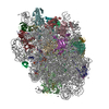 8camC  8cazC  8cepC  8ceuC  8cf1C  8cf8C  8cgdC  8cgiC  8cgjC 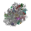 8cgkC 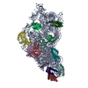 8cgrC 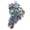 8cguC  8cgvC C: citing same article ( M: atomic model generated by this map |
|---|---|
| Similar structure data |

-Links
| EMDB pages | EMDB (EBI/PDBe) / EMDataResource |
|---|---|
| Related items in Molecule of the Month |


-Map
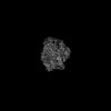
| File | Download / File: emd_16520.map.gz / Format: CCP4 / Size: 512 MB / Type: IMAGE STORED AS FLOATING POINT NUMBER (4 BYTES) | ||||||||||||||||||||||||||||||||||||
|---|---|---|---|---|---|---|---|---|---|---|---|---|---|---|---|---|---|---|---|---|---|---|---|---|---|---|---|---|---|---|---|---|---|---|---|---|---|
| Projections & slices | Image control
Images are generated by Spider. | ||||||||||||||||||||||||||||||||||||
| Voxel size | X=Y=Z: 0.762 Å | ||||||||||||||||||||||||||||||||||||
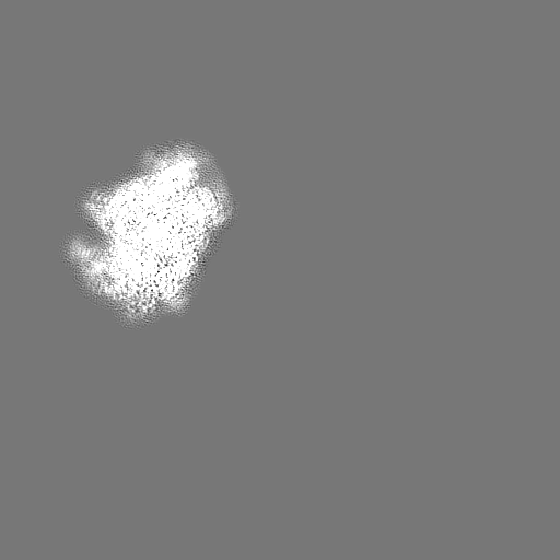
| Density |
| ||||||||||||||||||||||||||||||||||||
| Symmetry | Space group: 1 | ||||||||||||||||||||||||||||||||||||
| Details | EMDB XML:
|
 Z (Sec.)
 Y (Row.)
 X (Col.)


-Supplemental data
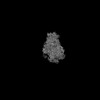
-Additional map: Sharpened map
| File | emd_16520_additional_1.map | ||||||||||||
|---|---|---|---|---|---|---|---|---|---|---|---|---|---|
| Annotation | Sharpened map | ||||||||||||
| Projections & Slices |
| ||||||||||||
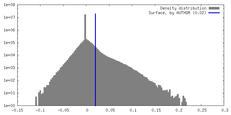
| Density Histograms |


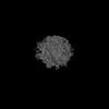
-Half map: #2
| File | emd_16520_half_map_1.map | ||||||||||||
|---|---|---|---|---|---|---|---|---|---|---|---|---|---|
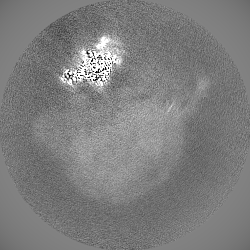
| Projections & Slices |
| ||||||||||||
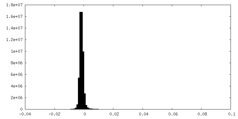
| Density Histograms |


-Half map: #1
| File | emd_16520_half_map_2.map | ||||||||||||
|---|---|---|---|---|---|---|---|---|---|---|---|---|---|
| Projections & Slices |
| ||||||||||||
| Density Histograms |


- Sample components
+Entire : Ribosomal small subunit head
+Supramolecule #1: Ribosomal small subunit head
+Macromolecule #1: 16S rRNA
+Macromolecule #2: Small ribosomal subunit protein uS3
+Macromolecule #3: Small ribosomal subunit protein uS5
+Macromolecule #4: 30S ribosomal protein S7
+Macromolecule #5: Small ribosomal subunit protein uS9
+Macromolecule #6: Small ribosomal subunit protein uS10
+Macromolecule #7: Small ribosomal subunit protein uS13
+Macromolecule #8: Small ribosomal subunit protein uS14
+Macromolecule #9: Small ribosomal subunit protein uS19
+Macromolecule #10: POTASSIUM ION
+Macromolecule #11: Omadacycline
+Macromolecule #12: SPECTINOMYCIN
+Macromolecule #13: MAGNESIUM ION
+Macromolecule #14: water

-Experimental details
-Structure determination
| Method | cryo EM |
|---|---|
 Processing Processing | single particle reconstruction |
| Aggregation state | particle |
-Sample preparation
| Buffer | pH: 7.5 Component:
| |||||||||||||||||||||
|---|---|---|---|---|---|---|---|---|---|---|---|---|---|---|---|---|---|---|---|---|---|---|
| Grid | Model: Quantifoil R1.2/1.3 / Material: GOLD / Mesh: 300 / Support film - Material: GRAPHENE | |||||||||||||||||||||
| Vitrification | Cryogen name: ETHANE / Chamber humidity: 100 % / Chamber temperature: 277.15 K |
- Electron microscopy
| Microscope | FEI TITAN KRIOS |
|---|---|
| Image recording | Film or detector model: GATAN K2 SUMMIT (4k x 4k) / Detector mode: COUNTING / Number real images: 24195 / Average exposure time: 4.0 sec. / Average electron dose: 44.0 e/Å2 |
| Electron beam | Acceleration voltage: 300 kV / Electron source:  FIELD EMISSION GUN FIELD EMISSION GUN |
| Electron optics | Illumination mode: FLOOD BEAM / Imaging mode: BRIGHT FIELD / Nominal defocus max: 1.6 µm / Nominal defocus min: 0.4 µm |
| Experimental equipment |  Model: Titan Krios / Image courtesy: FEI Company |
-Image processing
| Startup model | Type of model: PDB ENTRY PDB model - PDB ID: |
|---|---|
| Final reconstruction | Resolution.type: BY AUTHOR / Resolution: 2.06 Å / Resolution method: FSC 0.143 CUT-OFF / Number images used: 514855 |
| Initial angle assignment | Type: MAXIMUM LIKELIHOOD |
| Final angle assignment | Type: MAXIMUM LIKELIHOOD |

