 Movie
Movie Controller


+ Open data

- Basic information
| Entry | 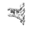 | |||||||||
|---|---|---|---|---|---|---|---|---|---|---|
| Title | Single-stranded Paranemic Crossover RNA Triangle (PXT) | |||||||||
 Map data Map data | ||||||||||
 Sample Sample |
| |||||||||
 Keywords Keywords | Cotranscriptional folding / RNA origami / RNA nanotechnology / Paranemic crossover / RNA | |||||||||
| Biological species | synthetic construct (others) | |||||||||
| Method | single particle reconstruction / cryo EM / Resolution: 5.39 Å | |||||||||
 Authors Authors | Sampedro N / McRae EKS / Andersen ES | |||||||||
| Funding support |  Denmark, 1 items Denmark, 1 items
| |||||||||
 Citation Citation | Journal: Small / Year: 2023 Title: An RNA Paranemic Crossover Triangle as A 3D Module for Cotranscriptional Nanoassembly. Authors: Néstor Sampedro Vallina / Ewan K S McRae / Cody Geary / Ebbe Sloth Andersen / Abstract: RNA nanotechnology takes advantage of structural modularity to build self-assembling nano-architectures with applications in medicine and synthetic biology. The use of paranemic motifs, that form ...RNA nanotechnology takes advantage of structural modularity to build self-assembling nano-architectures with applications in medicine and synthetic biology. The use of paranemic motifs, that form without unfolding existing secondary structure, allows for the creation of RNA nanostructures that are compatible with cotranscriptional folding in vitro and in vivo. In previous work, kissing-loop (KL) motifs have been widely used to design RNA nanostructures that fold cotranscriptionally. However, the paranemic crossover (PX) motif has not yet been explored for cotranscriptional RNA origami architectures and information about the structural geometry of the motif is unknown. Here, a six base pair-wide paranemic RNA interaction that arranges double helices in a perpendicular manner is introduced, allowing for the generation of a new and versatile building block: the paranemic-crossover triangle (PXT). The PXT is self-assembled by cotranscriptional folding and characterized by cryogenic electron microscopy, revealing for the first time an RNA PX interaction in high structural detail. The PXT is used as a building block for the construction of multimers that form filaments and rings and a duplicated PXT motif is used as a building block to self-assemble cubic structures, demonstrating the PXT as a rigid self-folding domain for the development of wireframe RNA origami architectures. | |||||||||
| History |
|
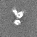
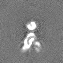
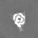
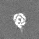
- Structure visualization
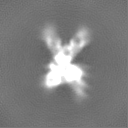
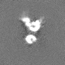
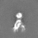
| Supplemental images |
|---|

- Downloads & links
-EMDB archive
| Map data | emd_16244.map.gz | 7.5 MB |  EMDB map data format EMDB map data format | |
|---|---|---|---|---|
| Header (meta data) | emd-16244-v30.xmlemd-16244.xml | 15.7 KB 15.7 KB | Display Display | EMDB header |
| Images |  emd_16244.png emd_16244.png | 26.7 KB | ||
| Filedesc metadata | emd-16244.cif.gz | 5 KB | ||
| Others | emd_16244_half_map_1.map.gzemd_16244_half_map_2.map.gz | 7.4 MB 7.4 MB | ||
| Archive directory |  http://ftp.pdbj.org/pub/emdb/structures/EMD-16244ftp://ftp.pdbj.org/pub/emdb/structures/EMD-16244 http://ftp.pdbj.org/pub/emdb/structures/EMD-16244ftp://ftp.pdbj.org/pub/emdb/structures/EMD-16244 | HTTPS FTP |
-Validation report
| Summary document | emd_16244_validation.pdf.gz | 664.8 KB | Display | EMDB validaton report |
|---|---|---|---|---|
| Full document | emd_16244_full_validation.pdf.gz | 664.4 KB | Display | |
| Data in XML | emd_16244_validation.xml.gz | 8.7 KB | Display | |
| Data in CIF | emd_16244_validation.cif.gz | 10 KB | Display | |
| Arichive directory | https://ftp.pdbj.org/pub/emdb/validation_reports/EMD-16244ftp://ftp.pdbj.org/pub/emdb/validation_reports/EMD-16244 | HTTPS FTP |
-Related structure data


-Links
| EMDB pages | EMDB (EBI/PDBe) / EMDataResource |
|---|
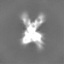
-Map
| File | Download / File: emd_16244.map.gz / Format: CCP4 / Size: 8 MB / Type: IMAGE STORED AS FLOATING POINT NUMBER (4 BYTES) | ||||||||||||||||||||
|---|---|---|---|---|---|---|---|---|---|---|---|---|---|---|---|---|---|---|---|---|---|
| Voxel size | X=Y=Z: 1.41 Å | ||||||||||||||||||||
| Density |
| ||||||||||||||||||||
| Symmetry | Space group: 1 | ||||||||||||||||||||
| Details | EMDB XML:
|
-Supplemental data
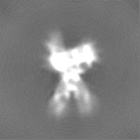
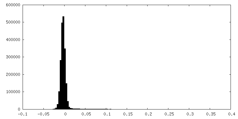
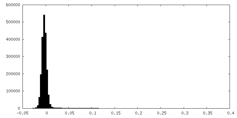
-Half map: Half map A
| File | emd_16244_half_map_1.map | ||||||||||||
|---|---|---|---|---|---|---|---|---|---|---|---|---|---|
| Annotation | Half_map_A | ||||||||||||
| Projections & Slices |
| ||||||||||||
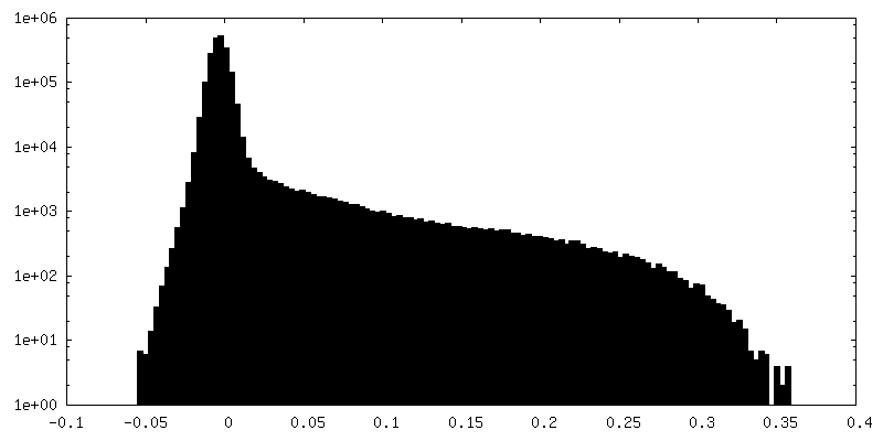
| Density Histograms |
 Z
Z Y
Y X
X
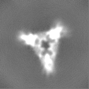

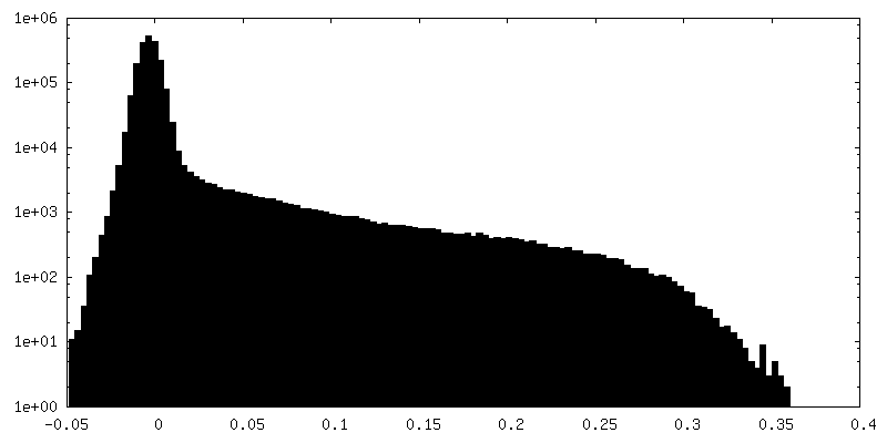
-Half map: Half map B
| File | emd_16244_half_map_2.map | ||||||||||||
|---|---|---|---|---|---|---|---|---|---|---|---|---|---|
| Annotation | Half_map_B | ||||||||||||
| Projections & Slices |
| ||||||||||||
| Density Histograms |


- Sample components
-Entire : Enzymatically synthesized and isothermally folded single stranded...
| Entire | Name: Enzymatically synthesized and isothermally folded single stranded RNA origami |
|---|---|
| Components |
|
-Supramolecule #1: Enzymatically synthesized and isothermally folded single stranded...
| Supramolecule | Name: Enzymatically synthesized and isothermally folded single stranded RNA origami type: complex / ID: 1 / Parent: 0 / Macromolecule list: all / Details: In vitro transcribed RNA purified by SEC. |
|---|---|
| Source (natural) | Organism: synthetic construct (others) |
| Molecular weight | Theoretical: 76.38 kDa/nm |
-Macromolecule #1: RNA Paranemic croosover triangle (PXT)
| Macromolecule | Name: RNA Paranemic croosover triangle (PXT) / type: rna / ID: 1 / Details: In vitro transcribed RNA / Number of copies: 1 |
|---|---|
| Source (natural) | Organism: synthetic construct (others) |
| Molecular weight | Theoretical: 76.393867 KDa |
| Sequence | String: GGAAUAUCGU CAUGGUGAUU CGUCACCAUG AGGCUAGAUC UCAUAUCUAG CGCUUUCGAG CGCUAGAGUC CUUAUCUAGC CGGUUUAUA CUUUCGAGUG UGAACCCGAU AUUCCGCGGA UCACUAUGAG UCGUUCGCGG CUCAUAGUCC GGCUCAAAGG A CAUCAUGG ...String: GGAAUAUCGU CAUGGUGAUU CGUCACCAUG AGGCUAGAUC UCAUAUCUAG CGCUUUCGAG CGCUAGAGUC CUUAUCUAGC CGGUUUAUA CUUUCGAGUG UGAACCCGAU AUUCCGCGGA UCACUAUGAG UCGUUCGCGG CUCAUAGUCC GGCUCAAAGG A CAUCAUGG CCUGUUCGCA GGUUGUGAUU AUGAGUGAGC CGGGUAAGGC AUACCGUUCG CGGUAUGUCU UACGAUCCGC |
-Experimental details
-Structure determination
| Method | cryo EM |
|---|---|
 Processing Processing | single particle reconstruction |
| Aggregation state | particle |
-Sample preparation
| Concentration | 3 mg/mL | ||||||||||||
|---|---|---|---|---|---|---|---|---|---|---|---|---|---|
| Buffer | pH: 7.8 Component:
Details: Filtered through 0.22 micron filter | ||||||||||||
| Vitrification | Cryogen name: ETHANE / Chamber humidity: 99 % / Chamber temperature: 294 K / Instrument: LEICA EM GP Details: 3 microliter droplet, 4 second delay before blotting, 6 second blot, 0 second delay before plunging.. | ||||||||||||
| Details | Sample was purified by size exclusion chromatography and concentrated in an Amicon spin concentrator. |
- Electron microscopy
| Microscope | FEI TITAN KRIOS |
|---|---|
| Specialist optics | Energy filter - Name: GIF Bioquantum / Energy filter - Slit width: 20 eV |
| Image recording | Film or detector model: GATAN K3 BIOQUANTUM (6k x 4k) / Average exposure time: 1.5 sec. / Average electron dose: 60.0 e/Å2 |
| Electron beam | Acceleration voltage: 300 kV / Electron source:  FIELD EMISSION GUN FIELD EMISSION GUN |
| Electron optics | C2 aperture diameter: 50.0 µm / Calibrated magnification: 130000 / Illumination mode: FLOOD BEAM / Imaging mode: BRIGHT FIELD / Cs: 2.7 mm / Nominal defocus max: 2.5 µm / Nominal defocus min: 0.75 µm |
| Sample stage | Specimen holder model: FEI TITAN KRIOS AUTOGRID HOLDER / Cooling holder cryogen: NITROGEN |
| Experimental equipment |  Model: Titan Krios / Image courtesy: FEI Company |
-Image processing
| Startup model | Type of model: NONE |
|---|---|
| Final reconstruction | Number classes used: 1 / Resolution.type: BY AUTHOR / Resolution: 5.39 Å / Resolution method: FSC 0.143 CUT-OFF / Software - Name: cryoSPARC (ver. 3.2.0) / Number images used: 437831 |
| Initial angle assignment | Type: MAXIMUM LIKELIHOOD |
| Final angle assignment | Type: MAXIMUM LIKELIHOOD |
-Atomic model buiding 1
| Refinement | Protocol: FLEXIBLE FIT |
|---|---|
| Output model |  PDB-8btz: |
