Journal: iScience / Year: 2023 Title: A nanobody recognizes a unique conserved epitope and potently neutralizes SARS-CoV-2 omicron variants. Authors: Naphak Modhiran / Simon Malte Lauer / Alberto A Amarilla / Peter Hewins / Sara Irene Lopes van den Broek / Yu Shang Low / Nazia Thakur / Benjamin Liang / Guillermo Valenzuela Nieto / James ...Authors: Naphak Modhiran / Simon Malte Lauer / Alberto A Amarilla / Peter Hewins / Sara Irene Lopes van den Broek / Yu Shang Low / Nazia Thakur / Benjamin Liang / Guillermo Valenzuela Nieto / James Jung / Devina Paramitha / Ariel Isaacs / Julian D J Sng / David Song / Jesper Tranekjær Jørgensen / Yorka Cheuquemilla / Jörg Bürger / Ida Vang Andersen / Johanna Himelreichs / Ronald Jara / Ronan MacLoughlin / Zaray Miranda-Chacon / Pedro Chana-Cuevas / Vasko Kramer / Christian Spahn / Thorsten Mielke / Alexander A Khromykh / Trent Munro / Martina L Jones / Paul R Young / Keith Chappell / Dalan Bailey / Andreas Kjaer / Matthias Manfred Herth / Kellie Ann Jurado / David Schwefel / Alejandro Rojas-Fernandez / Daniel Watterson / Abstract: The severe acute respiratory syndrome coronavirus 2 (SARS-CoV2) Omicron variant sub-lineages spread rapidly worldwide, mostly due to their immune-evasive properties. This has put a significant part ...The severe acute respiratory syndrome coronavirus 2 (SARS-CoV2) Omicron variant sub-lineages spread rapidly worldwide, mostly due to their immune-evasive properties. This has put a significant part of the population at risk for severe disease and underscores the need for effective anti-SARS-CoV-2 agents against emergent strains in vulnerable patients. Camelid nanobodies are attractive therapeutic candidates due to their high stability, ease of large-scale production, and potential for delivery via inhalation. Here, we characterize the receptor binding domain (RBD)-specific nanobody W25 and show superior neutralization activity toward Omicron sub-lineages in comparison to all other SARS-CoV2 variants. Structure analysis of W25 in complex with the SARS-CoV2 spike glycoprotein shows that W25 engages an RBD epitope not covered by any of the antibodies previously approved for emergency use. evaluation of W25 prophylactic and therapeutic treatments across multiple SARS-CoV-2 variant infection models, together with W25 biodistribution analysis in mice, demonstrates favorable pre-clinical properties. Together, these data endorse W25 for further clinical development.
Film or detector model: GATAN K3 (6k x 4k) / Number grids imaged: 1 / Number real images: 9125 / Average exposure time: 3.3 sec. / Average electron dose: 40.0 e/Å2
Electron beam
Acceleration voltage: 300 kV / Electron source: FIELD EMISSION GUN
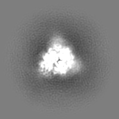
Number classes used: 1 / Resolution.type: BY AUTHOR / Resolution: 6.04 Å / Resolution method: FSC 0.143 CUT-OFF / Software - Name: cryoSPARC (ver. 3.3.1) / Number images used: 32512
Initial angle assignment
Type: MAXIMUM LIKELIHOOD / Software - Name: cryoSPARC (ver. 3.3.1)
Final angle assignment
Type: MAXIMUM LIKELIHOOD / Software - Name: cryoSPARC (ver. 3.3.1)
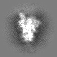
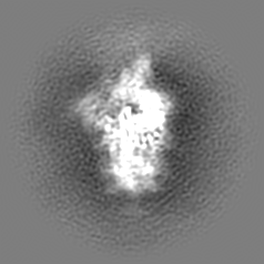
Final 3D classification
Number classes: 3 / Software - Name: cryoSPARC (ver. 3.3.1)
-
Atomic model buiding 1
Refinement
Space: REAL / Protocol: OTHER
Output model
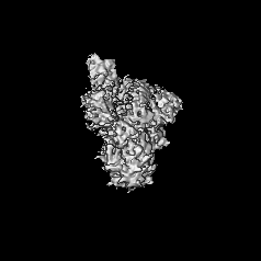
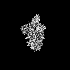
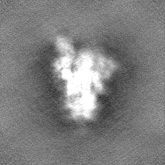
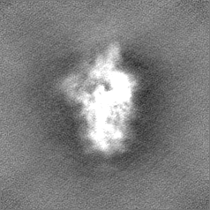
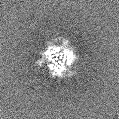
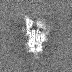
PDB-8bgg: Cryo-EM structure of SARS-CoV-2 spike (Omicron BA.1 variant) in complex with nanobody W25 (map 5, focus refinement on RBD, W25 and adjacent NTD)
+
About Yorodumi
-
News
-
Feb 9, 2022. New format data for meta-information of EMDB entries
New format data for meta-information of EMDB entries
Version 3 of the EMDB header file is now the official format.
The previous official version 1.9 will be removed from the archive.
In the structure databanks used in Yorodumi, some data are registered as the other names, "COVID-19 virus" and "2019-nCoV". Here are the details of the virus and the list of structure data.
Jan 31, 2019. EMDB accession codes are about to change! (news from PDBe EMDB page)
EMDB accession codes are about to change! (news from PDBe EMDB page)
The allocation of 4 digits for EMDB accession codes will soon come to an end. Whilst these codes will remain in use, new EMDB accession codes will include an additional digit and will expand incrementally as the available range of codes is exhausted. The current 4-digit format prefixed with “EMD-” (i.e. EMD-XXXX) will advance to a 5-digit format (i.e. EMD-XXXXX), and so on. It is currently estimated that the 4-digit codes will be depleted around Spring 2019, at which point the 5-digit format will come into force.
The EM Navigator/Yorodumi systems omit the EMD- prefix.
Related info.:Q: What is EMD? / ID/Accession-code notation in Yorodumi/EM Navigator
Yorodumi is a browser for structure data from EMDB, PDB, SASBDB, etc.
This page is also the successor to EM Navigator detail page, and also detail information page/front-end page for Omokage search.
The word "yorodu" (or yorozu) is an old Japanese word meaning "ten thousand". "mi" (miru) is to see.
Related info.:EMDB / PDB / SASBDB / Comparison of 3 databanks / Yorodumi Search / Aug 31, 2016. New EM Navigator & Yorodumi / Yorodumi Papers / Jmol/JSmol / Function and homology information / Changes in new EM Navigator and Yorodumi
 Movie
Movie Controller

 Yorodumi
 Open data
 Basic information
 Map data
 Sample
 Keywords
 Function and homology information
 Severe acute respiratory syndrome coronavirus 2 /
 Authors
 Germany, 1 items
  Citation


 Structure visualization
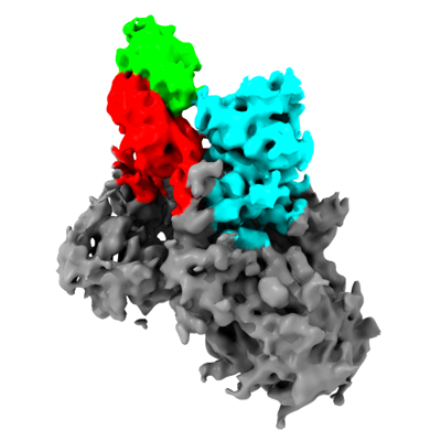
 Downloads & links
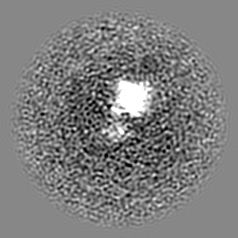
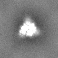
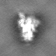
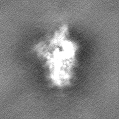
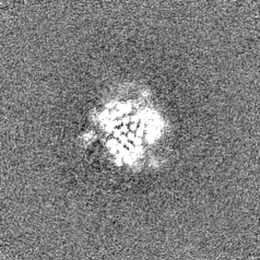
 emd_16030.png
 http://ftp.pdbj.org/pub/emdb/structures/EMD-16030


 Z (Sec.)
 Y (Row.)
 X (Col.)


 Sample components
 Homo sapiens (human)

 Processing
 Electron microscopy
 FIELD EMISSION GUN