Movie
Movie Controller

[English] 日本語
 Yorodumi
- EMDB-15691: Cryo-EM structure of heme A synthase trimer from Aquifex aeolicus -
+ Open data

- Basic information
| Entry | 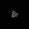 | |||||||||
|---|---|---|---|---|---|---|---|---|---|---|
| Title | Cryo-EM structure of heme A synthase trimer from Aquifex aeolicus | |||||||||
 Map data Map data | ||||||||||
 Sample Sample |
| |||||||||
 Keywords Keywords | Heme A synthase / Oligomerization / Function / Heme A / MEMBRANE PROTEIN | |||||||||
| Function / homology | COX15/CtaA family / Cytochrome oxidase assembly protein / oxidoreductase activity, acting on NAD(P)H, heme protein as acceptor / heme A biosynthetic process / membrane / Heme O oxygenase Function and homology information Function and homology information | |||||||||
| Biological species |   Aquifex aeolicus VF5 (bacteria) Aquifex aeolicus VF5 (bacteria) | |||||||||
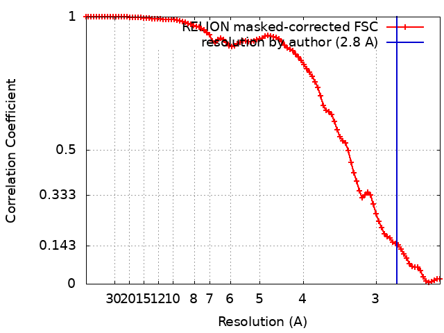
| Method | single particle reconstruction / cryo EM / Resolution: 2.8 Å | |||||||||
 Authors Authors | Hui Z / Guoliang Z | |||||||||
| Funding support |  China, 2 items China, 2 items
| |||||||||
 Citation Citation | Journal: To Be Published Title: Cryo-EM structure of heme A synthase trimer from Aquifex aeolicus Authors: Hui Z / Guoliang Z | |||||||||
| History |
|
- Structure visualization
| Supplemental images |
|---|

- Downloads & links
-EMDB archive
| Map data | emd_15691.map.gz | 4.8 MB | EMDB map data format | |
|---|---|---|---|---|
| Header (meta data) | emd-15691-v30.xmlemd-15691.xml | 15.3 KB 15.3 KB | Display Display | EMDB header |
| FSC (resolution estimation) | emd_15691_fsc.xml | 9.1 KB | Display | FSC data file |
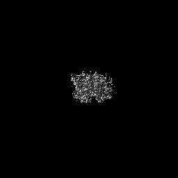
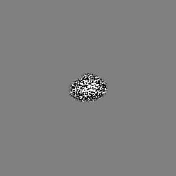
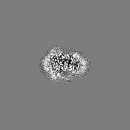
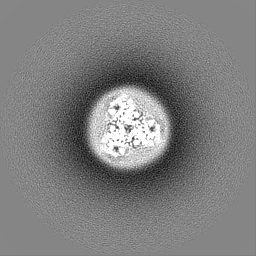
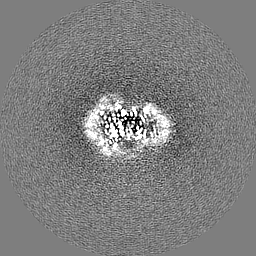
| Images | 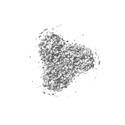 emd_15691.png emd_15691.png | 53.2 KB | ||
| Masks | emd_15691_msk_1.map | 64 MB | Mask map | |
| Others | emd_15691_half_map_1.map.gzemd_15691_half_map_2.map.gz | 49.7 MB 49.7 MB | ||
| Archive directory |  http://ftp.pdbj.org/pub/emdb/structures/EMD-15691ftp://ftp.pdbj.org/pub/emdb/structures/EMD-15691 http://ftp.pdbj.org/pub/emdb/structures/EMD-15691ftp://ftp.pdbj.org/pub/emdb/structures/EMD-15691 | HTTPS FTP |
-Validation report
| Summary document | emd_15691_validation.pdf.gz | 677.1 KB | Display | EMDB validaton report |
|---|---|---|---|---|
| Full document | emd_15691_full_validation.pdf.gz | 676.7 KB | Display | |
| Data in XML | emd_15691_validation.xml.gz | 15.9 KB | Display | |
| Data in CIF | emd_15691_validation.cif.gz | 21 KB | Display | |
| Arichive directory | https://ftp.pdbj.org/pub/emdb/validation_reports/EMD-15691ftp://ftp.pdbj.org/pub/emdb/validation_reports/EMD-15691 | HTTPS FTP |
-Related structure data
| Related structure data | 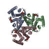 8aw5MC M: atomic model generated by this map C: citing same article ( |
|---|---|
| Similar structure data |
-Links
| EMDB pages | EMDB (EBI/PDBe) / EMDataResource |
|---|
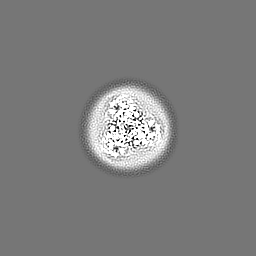
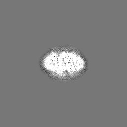
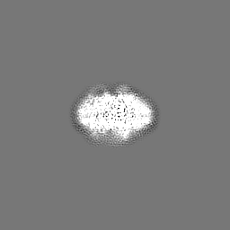
-Map
| File | Download / File: emd_15691.map.gz / Format: CCP4 / Size: 64 MB / Type: IMAGE STORED AS FLOATING POINT NUMBER (4 BYTES) | ||||||||||||||||||||||||||||||||||||
|---|---|---|---|---|---|---|---|---|---|---|---|---|---|---|---|---|---|---|---|---|---|---|---|---|---|---|---|---|---|---|---|---|---|---|---|---|---|
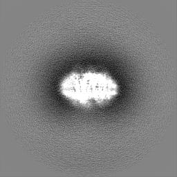
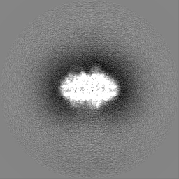
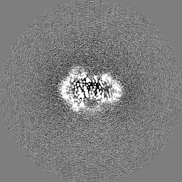
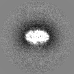
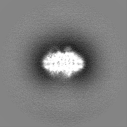
| Projections & slices | Image control
Images are generated by Spider. | ||||||||||||||||||||||||||||||||||||
| Voxel size | X=Y=Z: 1.23 Å | ||||||||||||||||||||||||||||||||||||
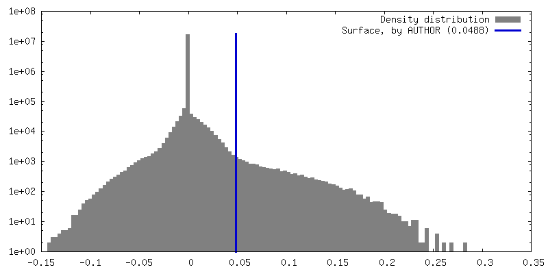
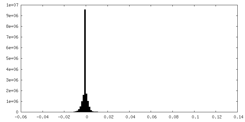
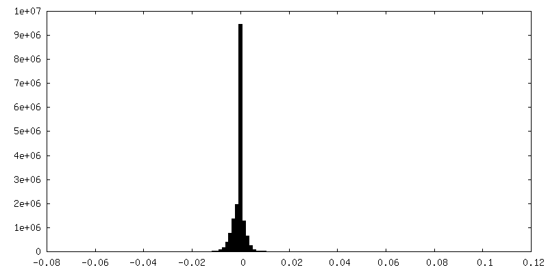
| Density |
| ||||||||||||||||||||||||||||||||||||
| Symmetry | Space group: 1 | ||||||||||||||||||||||||||||||||||||
| Details | EMDB XML:
|
 Z (Sec.)
 Y (Row.)
 X (Col.)


-Supplemental data
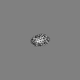
-Mask #1
| File | emd_15691_msk_1.map | ||||||||||||
|---|---|---|---|---|---|---|---|---|---|---|---|---|---|
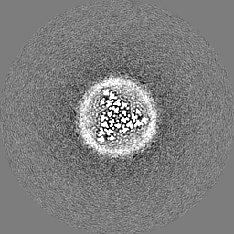
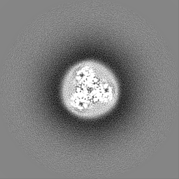
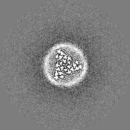
| Projections & Slices |
| ||||||||||||
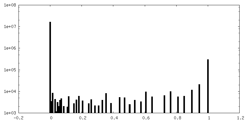
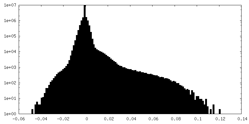
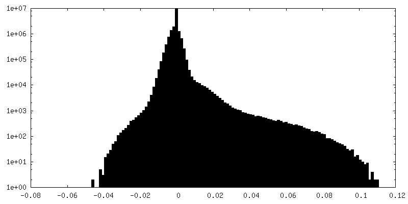
| Density Histograms |


-Half map: #2
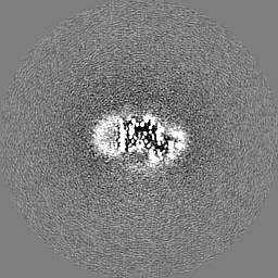
| File | emd_15691_half_map_1.map | ||||||||||||
|---|---|---|---|---|---|---|---|---|---|---|---|---|---|
| Projections & Slices |
| ||||||||||||
| Density Histograms |

-Half map: #1
| File | emd_15691_half_map_2.map | ||||||||||||
|---|---|---|---|---|---|---|---|---|---|---|---|---|---|
| Projections & Slices |
| ||||||||||||
| Density Histograms |
- Sample components
-Entire : Heme A synthase with cofactor Heme B
| Entire | Name: Heme A synthase with cofactor Heme B |
|---|---|
| Components |
|
-Supramolecule #1: Heme A synthase with cofactor Heme B
| Supramolecule | Name: Heme A synthase with cofactor Heme B / type: complex / ID: 1 / Parent: 0 / Macromolecule list: #1 |
|---|---|
| Source (natural) | Organism: Aquifex aeolicus VF5 (bacteria) |
-Macromolecule #1: Heme O oxygenase
| Macromolecule | Name: Heme O oxygenase / type: protein_or_peptide / ID: 1 / Number of copies: 3 / Enantiomer: LEVO |
|---|---|
| Source (natural) | Organism: Aquifex aeolicus VF5 (bacteria) / Strain: VF5 |
| Molecular weight | Theoretical: 34.984523 KDa |
| Recombinant expression | Organism: |
| Sequence | String: GMNTNLKSSP LKTLVLASVV LTYVLMVFGG IVTSTGSGLG CPDWPLCHGQ LLPFQLKEQI PTPPAPVVAP TPLQPWIEQT HRILGGITG IVLLATLFYA FKRGTSFVKK ALVFIFIALI LEALLGMRVV ITEAPLLREL LHYVYTSAHL ILSVFILSTI T ITYYYVKF ...String: GMNTNLKSSP LKTLVLASVV LTYVLMVFGG IVTSTGSGLG CPDWPLCHGQ LLPFQLKEQI PTPPAPVVAP TPLQPWIEQT HRILGGITG IVLLATLFYA FKRGTSFVKK ALVFIFIALI LEALLGMRVV ITEAPLLREL LHYVYTSAHL ILSVFILSTI T ITYYYVKF FGERPKEYIP YADALYVATM FQILLGIFVR YVKALEYNQF VYYLHITYAG FLVILSLFIM FKEFNKYSLI TF LLMTAQI LAGVATVISG FFLPYLFLHI AIGFFIVLWV SYLVAPSVLK TYTEFRGELA RNSSAWSHPQ FEK UniProtKB: Heme O oxygenase |
-Macromolecule #2: PROTOPORPHYRIN IX CONTAINING FE
| Macromolecule | Name: PROTOPORPHYRIN IX CONTAINING FE / type: ligand / ID: 2 / Number of copies: 3 / Formula: HEM |
|---|---|
| Molecular weight | Theoretical: 616.487 Da |
| Chemical component information |  ChemComp-HEM: |
-Macromolecule #3: (2S)-3-(hexadecanoyloxy)-2-[(9Z)-octadec-9-enoyloxy]propyl 2-(tri...
| Macromolecule | Name: (2S)-3-(hexadecanoyloxy)-2-[(9Z)-octadec-9-enoyloxy]propyl 2-(trimethylammonio)ethyl phosphate type: ligand / ID: 3 / Number of copies: 9 / Formula: POV |
|---|---|
| Molecular weight | Theoretical: 760.076 Da |
| Chemical component information |  ChemComp-POV: |
-Experimental details
-Structure determination
| Method | cryo EM |
|---|---|
 Processing Processing | single particle reconstruction |
| Aggregation state | 3D array |
-Sample preparation
| Concentration | 3.5 mg/mL | |||||||||
|---|---|---|---|---|---|---|---|---|---|---|
| Buffer | pH: 7.4 Component:
| |||||||||
| Grid | Model: Homemade / Material: GOLD / Mesh: 300 / Pretreatment - Type: GLOW DISCHARGE / Pretreatment - Time: 60 sec. / Pretreatment - Atmosphere: OTHER | |||||||||
| Vitrification | Cryogen name: ETHANE / Chamber humidity: 100 % / Chamber temperature: 277.15 K |
- Electron microscopy
| Microscope | FEI TITAN KRIOS |
|---|---|
| Specialist optics | Energy filter - Slit width: 20 eV |
| Image recording | Film or detector model: GATAN K2 SUMMIT (4k x 4k) / Detector mode: SUPER-RESOLUTION / Average electron dose: 70.0 e/Å2 |
| Electron beam | Acceleration voltage: 300 kV / Electron source: OTHER |
| Electron optics | Calibrated defocus min: 1.2 µm / Illumination mode: FLOOD BEAM / Imaging mode: BRIGHT FIELD / Nominal defocus max: 2.2 µm / Nominal defocus min: 1.1 µm / Nominal magnification: 165000 |
| Sample stage | Cooling holder cryogen: NITROGEN |
| Experimental equipment |  Model: Titan Krios / Image courtesy: FEI Company |