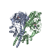
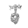
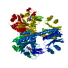
+ Open data
- Basic information
| Entry |  | |||||||||
|---|---|---|---|---|---|---|---|---|---|---|
| Title | Cryo-EM structure of DrBphP in Pr state | |||||||||
 Map data Map data | ||||||||||
 Sample Sample |
| |||||||||
 Keywords Keywords | KINASE / PHOTOSENSOR / TRANSFERASE / PHYTOCHROME | |||||||||
| Function / homology |  Function and homology information Function and homology informationosmosensory signaling via phosphorelay pathway / detection of visible light / phosphorelay response regulator activity / protein kinase activator activity / histidine kinase / phosphorelay sensor kinase activity / phosphorelay signal transduction system / photoreceptor activity / regulation of DNA-templated transcription / ATP binding ...osmosensory signaling via phosphorelay pathway / detection of visible light / phosphorelay response regulator activity / protein kinase activator activity / histidine kinase / phosphorelay sensor kinase activity / phosphorelay signal transduction system / photoreceptor activity / regulation of DNA-templated transcription / ATP binding / identical protein binding / metal ion binding Similarity search - Function | |||||||||
| Biological species |  Deinococcus radiodurans R1 (radioresistant) Deinococcus radiodurans R1 (radioresistant) | |||||||||
| Method | single particle reconstruction / cryo EM / Resolution: 3.62 Å | |||||||||
 Authors Authors | Wahlgren WY / Takala H / Westenhoff S | |||||||||
| Funding support | European Union,  Finland, 2 items Finland, 2 items
| |||||||||
 Citation Citation |  Journal: Nat Commun / Year: 2022 Journal: Nat Commun / Year: 2022Title: Structural mechanism of signal transduction in a phytochrome histidine kinase. Authors: Weixiao Yuan Wahlgren / Elin Claesson / Iida Tuure / Sergio Trillo-Muyo / Szabolcs Bódizs / Janne A Ihalainen / Heikki Takala / Sebastian Westenhoff /   Abstract: Phytochrome proteins detect red/far-red light to guide the growth, motion, development and reproduction in plants, fungi, and bacteria. Bacterial phytochromes commonly function as an entrance signal ...Phytochrome proteins detect red/far-red light to guide the growth, motion, development and reproduction in plants, fungi, and bacteria. Bacterial phytochromes commonly function as an entrance signal in two-component sensory systems. Despite the availability of three-dimensional structures of phytochromes and other two-component proteins, the conformational changes, which lead to activation of the protein, are not understood. We reveal cryo electron microscopy structures of the complete phytochrome from Deinoccocus radiodurans in its resting and photoactivated states at 3.6 Å and 3.5 Å resolution, respectively. Upon photoactivation, the photosensory core module hardly changes its tertiary domain arrangement, but the connector helices between the photosensory and the histidine kinase modules open up like a zipper, causing asymmetry and disorder in the effector domains. The structures provide a framework for atom-scale understanding of signaling in phytochromes, visualize allosteric communication over several nanometers, and suggest that disorder in the dimeric arrangement of the effector domains is important for phosphatase activity in a two-component system. The results have implications for the development of optogenetic applications. | |||||||||
| History |
|
- Structure visualization
| Supplemental images |
|---|
- Downloads & links
-EMDB archive
| Map data |  emd_15685.map.gz emd_15685.map.gz | 263.7 MB |  EMDB map data format EMDB map data format | |
|---|---|---|---|---|
| Header (meta data) |  emd-15685-v30.xml emd-15685-v30.xml emd-15685.xml emd-15685.xml | 16.3 KB 16.3 KB | Display Display |  EMDB header EMDB header |
| FSC (resolution estimation) |  emd_15685_fsc.xml emd_15685_fsc.xml | 14.8 KB | Display |  FSC data file FSC data file |
| Images |  emd_15685.png emd_15685.png | 57.9 KB | ||
| Masks |  emd_15685_msk_1.map emd_15685_msk_1.map | 343 MB |  Mask map Mask map | |
| Filedesc metadata |  emd-15685.cif.gz emd-15685.cif.gz | 6.2 KB | ||
| Others |  emd_15685_half_map_1.map.gz emd_15685_half_map_1.map.gz emd_15685_half_map_2.map.gz emd_15685_half_map_2.map.gz | 258.6 MB 258.6 MB | ||
| Archive directory |  http://ftp.pdbj.org/pub/emdb/structures/EMD-15685 http://ftp.pdbj.org/pub/emdb/structures/EMD-15685 ftp://ftp.pdbj.org/pub/emdb/structures/EMD-15685 ftp://ftp.pdbj.org/pub/emdb/structures/EMD-15685 | HTTPS FTP |
-Validation report
| Summary document |  emd_15685_validation.pdf.gz emd_15685_validation.pdf.gz | 1 MB | Display |  EMDB validaton report EMDB validaton report |
|---|---|---|---|---|
| Full document |  emd_15685_full_validation.pdf.gz emd_15685_full_validation.pdf.gz | 1 MB | Display | |
| Data in XML |  emd_15685_validation.xml.gz emd_15685_validation.xml.gz | 24 KB | Display | |
| Data in CIF |  emd_15685_validation.cif.gz emd_15685_validation.cif.gz | 31.2 KB | Display | |
| Arichive directory |  https://ftp.pdbj.org/pub/emdb/validation_reports/EMD-15685 https://ftp.pdbj.org/pub/emdb/validation_reports/EMD-15685 ftp://ftp.pdbj.org/pub/emdb/validation_reports/EMD-15685 ftp://ftp.pdbj.org/pub/emdb/validation_reports/EMD-15685 | HTTPS FTP |
-Related structure data
| Related structure data |  8avwMC  8avvC  8avxC M: atomic model generated by this map C: citing same article ( |
|---|---|
| Similar structure data | Similarity search - Function & homology  F&H Search F&H Search |
- Links
Links
| EMDB pages |  EMDB (EBI/PDBe) / EMDB (EBI/PDBe) /  EMDataResource EMDataResource |
|---|---|
| Related items in Molecule of the Month |
- Map
Map
| File |  Download / File: emd_15685.map.gz / Format: CCP4 / Size: 343 MB / Type: IMAGE STORED AS FLOATING POINT NUMBER (4 BYTES) Download / File: emd_15685.map.gz / Format: CCP4 / Size: 343 MB / Type: IMAGE STORED AS FLOATING POINT NUMBER (4 BYTES) | ||||||||||||||||||||||||||||||||||||
|---|---|---|---|---|---|---|---|---|---|---|---|---|---|---|---|---|---|---|---|---|---|---|---|---|---|---|---|---|---|---|---|---|---|---|---|---|---|
| Projections & slices | Image control
Images are generated by Spider. | ||||||||||||||||||||||||||||||||||||
| Voxel size | X=Y=Z: 0.8617 Å | ||||||||||||||||||||||||||||||||||||
| Density |
| ||||||||||||||||||||||||||||||||||||
| Symmetry | Space group: 1 | ||||||||||||||||||||||||||||||||||||
| Details | EMDB XML:
|
-Supplemental data
-Mask #1
| File |  emd_15685_msk_1.map emd_15685_msk_1.map | ||||||||||||
|---|---|---|---|---|---|---|---|---|---|---|---|---|---|
| Projections & Slices |
| ||||||||||||
| Density Histograms |
-Half map: #1
| File | emd_15685_half_map_1.map | ||||||||||||
|---|---|---|---|---|---|---|---|---|---|---|---|---|---|
| Projections & Slices |
| ||||||||||||
| Density Histograms |
-Half map: #2
| File | emd_15685_half_map_2.map | ||||||||||||
|---|---|---|---|---|---|---|---|---|---|---|---|---|---|
| Projections & Slices |
| ||||||||||||
| Density Histograms |
- Sample components
-Entire : Full-length bacteriophytochrome fused to its response regulator f...
| Entire | Name: Full-length bacteriophytochrome fused to its response regulator from Deinococcus radiodurans. |
|---|---|
| Components |
|
-Supramolecule #1: Full-length bacteriophytochrome fused to its response regulator f...
| Supramolecule | Name: Full-length bacteriophytochrome fused to its response regulator from Deinococcus radiodurans. type: complex / ID: 1 / Parent: 0 / Macromolecule list: #1 |
|---|---|
| Source (natural) | Organism:  Deinococcus radiodurans R1 (radioresistant) Deinococcus radiodurans R1 (radioresistant) |
| Molecular weight | Theoretical: 101 KDa |
-Macromolecule #1: Bacteriophytochrome,Response regulator
| Macromolecule | Name: Bacteriophytochrome,Response regulator / type: protein_or_peptide / ID: 1 Details: Sample is a fusion of Deinococcus radiodurans DrBphP and its response regulator DrRR.,Sample is a fusion of Deinococcus radiodurans DrBphP and its response regulator DrRR. Number of copies: 2 / Enantiomer: LEVO / EC number: histidine kinase |
|---|---|
| Source (natural) | Organism:  Deinococcus radiodurans R1 (radioresistant) Deinococcus radiodurans R1 (radioresistant)Strain: ATCC 13939 / DSM 20539 / JCM 16871 / LMG 4051 / NBRC 15346 / NCIMB 9279 / R1 / VKM B-1422 |
| Molecular weight | Theoretical: 101.579164 KDa |
| Recombinant expression | Organism:  |
| Sequence | String: MASMTGGQQM GRGSMSRDPL PFFPPLYLGG PEITTENCER EPIHIPGSIQ PHGALLTADG HSGEVLQMSL NAATFLGQEP TVLRGQTLA ALLPEQWPAL QAALPPGCPD ALQYRATLDW PAAGHLSLTV HRVGELLILE FEPTEAWDST GPHALRNAMF A LESAPNLR ...String: MASMTGGQQM GRGSMSRDPL PFFPPLYLGG PEITTENCER EPIHIPGSIQ PHGALLTADG HSGEVLQMSL NAATFLGQEP TVLRGQTLA ALLPEQWPAL QAALPPGCPD ALQYRATLDW PAAGHLSLTV HRVGELLILE FEPTEAWDST GPHALRNAMF A LESAPNLR ALAEVATQTV RELTGFDRVM LYKFAPDATG EVIAEARREG LHAFLGHRFP ASDIPAQARA LYTRHLLRLT AD TRAAAVP LDPVLNPQTN APTPLGGAVL RATSPMHMQY LRNMGVGSSL SVSVVVGGQL WGLIACHHQT PYVLPPDLRT TLE YLGRLL SLQVQVKEAA DVAAFRQSLR EHHARVALAA AHSLSPHDTL SDPALDLLGL MRAGGLILRF EGRWQTLGEV PPAP AVDAL LAWLETQPGA LVQTDALGQL WPAGADLAPS AAGLLAISVG EGWSECLVWL RPELRLEVAW GGATPDQAKD DLGPR HSFD TYLEEKRGYA EPWHPGEIEE AQDLRDTLTG ALGERLSVIR DLNRALTQSN AEWRQYGFVI SHHMQEPVRL ISQFAE LLT RQPRAQDGSP DSPQTERITG FLLRETSRLR SLTQDLHTYT ALLSAPPPVR RPTPLGRVVD DVLQDLEPRI ADTGASI EV APELPVIAAD AGLLRDLLLH LIGNALTFGG PEPRIAVRTE RQGAGWSIAV SDQGAGIAPE YQERIFLLFQ RLGSLDEA L GNGLGLPLCR KIAELHGGTL TVESAPGEGS TFRCWLPDAG PLPGAADAAS SAGGSAGSAG MPERASVPLR LLLVEDNAA DIFLMEMALE YSSVHTELLV ARDGLEALEL LEQAKTGGPF PDLILLDLNM PRVDGFELLQ ALRADPHLAH LPAIVLTTSN DPSDVKRAY ALQANSYLTK PSTLEDFLQL IERLTAYWFG TAAIPQTYQP QLEHHHHHH UniProtKB: Bacteriophytochrome, Response regulator |
-Macromolecule #2: 3-[2-[(Z)-[3-(2-carboxyethyl)-5-[(Z)-(4-ethenyl-3-methyl-5-oxidan...
| Macromolecule | Name: 3-[2-[(Z)-[3-(2-carboxyethyl)-5-[(Z)-(4-ethenyl-3-methyl-5-oxidanylidene-pyrrol-2-ylidene)methyl]-4-methyl-pyrrol-1-ium -2-ylidene]methyl]-5-[(Z)-[(3E)-3-ethylidene-4-methyl-5-oxidanylidene- ...Name: 3-[2-[(Z)-[3-(2-carboxyethyl)-5-[(Z)-(4-ethenyl-3-methyl-5-oxidanylidene-pyrrol-2-ylidene)methyl]-4-methyl-pyrrol-1-ium -2-ylidene]methyl]-5-[(Z)-[(3E)-3-ethylidene-4-methyl-5-oxidanylidene-pyrrolidin-2-ylidene]methyl]-4-methyl-1H-pyrrol-3- yl]propanoic acid type: ligand / ID: 2 / Number of copies: 2 / Formula: LBV |
|---|---|
| Molecular weight | Theoretical: 585.67 Da |
-Experimental details
-Structure determination
| Method | cryo EM |
|---|---|
 Processing Processing | single particle reconstruction |
| Aggregation state | particle |
- Sample preparation
| Buffer | pH: 8 |
|---|---|
| Vitrification | Cryogen name: ETHANE |
- Electron microscopy
| Microscope | FEI TITAN KRIOS |
|---|---|
| Image recording | Film or detector model: GATAN K3 BIOQUANTUM (6k x 4k) / Average electron dose: 48.3 e/Å2 |
| Electron beam | Acceleration voltage: 300 kV / Electron source:  FIELD EMISSION GUN FIELD EMISSION GUN |
| Electron optics | Illumination mode: FLOOD BEAM / Imaging mode: BRIGHT FIELD / Nominal defocus max: 2.6 µm / Nominal defocus min: 0.6 µm |
| Experimental equipment |  Model: Titan Krios / Image courtesy: FEI Company |
 Movie
Movie Controller


 Z (Sec.)
 Y (Row.)
 X (Col.)


