photosystem / assembly factor / membrane protein / PHOTOSYNTHESIS
Function / homology
Function and homology information
plasma membrane-derived thylakoid photosystem II / thylakoid lumen / photosystem II assembly / thylakoid / oxygen evolving activity / photosystem II reaction center / photosystem II / photosystem I reaction center / photosystem I / photosynthetic electron transport in photosystem I ...plasma membrane-derived thylakoid photosystem II / thylakoid lumen / photosystem II assembly / thylakoid / oxygen evolving activity / photosystem II reaction center / photosystem II / photosystem I reaction center / photosystem I / photosynthetic electron transport in photosystem I / photosynthetic electron transport chain / oxidoreductase activity, acting on diphenols and related substances as donors, oxygen as acceptor / photosystem I / response to herbicide / photosystem II / plasma membrane-derived thylakoid membrane / photosynthetic electron transport in photosystem II / chlorophyll binding / photosynthesis / 4 iron, 4 sulfur cluster binding / electron transfer activity / oxidoreductase activity / iron ion binding / heme binding / magnesium ion binding / metal ion binding / identical protein binding / plasma membrane Similarity search - Function
Photosynthesis system II assembly factor Ycf48/Hcf136 / Photosynthesis system II assembly factor Ycf48/Hcf136-like domain / Photosynthesis system II assembly factor YCF48 / Photosystem I reaction center subunit PsaK / Photosystem I reaction centre subunit PsaK / Photosystem I reaction centre subunit PsaK superfamily / Photosystem I PsaM, reaction centre superfamily / Photosystem I psaG and psaK proteins signature. / Photosystem I PsaM, reaction centre / Photosystem I protein M (PsaM) ...Photosynthesis system II assembly factor Ycf48/Hcf136 / Photosynthesis system II assembly factor Ycf48/Hcf136-like domain / Photosynthesis system II assembly factor YCF48 / Photosystem I reaction center subunit PsaK / Photosystem I reaction centre subunit PsaK / Photosystem I reaction centre subunit PsaK superfamily / Photosystem I PsaM, reaction centre superfamily / Photosystem I psaG and psaK proteins signature. / Photosystem I PsaM, reaction centre / Photosystem I protein M (PsaM) / Photosystem I reaction center subunit V/PsaK / Photosystem I psaG / psaK / Photosystem II PsbI / Photosystem II PsbI superfamily / Photosystem II reaction centre I protein (PSII 4.8 kDa protein) / Photosystem II protein D1 / Photosystem II D2 protein / Photosystem II cytochrome b559, conserved site / Photosystem II cytochrome b559, alpha subunit / Photosystem II cytochrome b559, beta subunit / Photosystem II cytochrome b559, N-terminal / Photosystem II cytochrome b559, alpha subunit, lumenal region / Photosystem II cytochrome b559, alpha subunit superfamily / Cytochrome b559, alpha (gene psbE) and beta (gene psbF)subunits / Lumenal portion of Cytochrome b559, alpha (gene psbE) subunit / Cytochrome b559 subunits heme-binding site signature. / : / Photosystem I PsaL, reaction centre subunit XI / Photosystem I, reaction centre subunit XI / Photosystem I PsaL, reaction centre subunit XI superfamily / Photosystem I reaction centre subunit XI / Photosystem I reaction centre subunit VIII / Photosystem I reaction centre subunit VIII / Photosystem I reaction centre subunit VIII superfamily / Photosystem I PsaF, reaction centre subunit III / Photosystem I PsaF, reaction centre subunit III superfamily / Photosystem I reaction centre subunit III / Photosystem I PsaD / Photosystem I, reaction centre subunit PsaD superfamily / PsaD / Photosystem I PsaE, reaction centre subunit IV / Photosystem I reaction centre subunit IV / PsaE / Photosystem I PsaJ, reaction centre subunit IX superfamily / Photosystem I PsaJ, reaction centre subunit IX / Photosystem I reaction centre subunit IX / PsaJ / Photosystem I PsaA / Photosystem I protein PsaC / Photosystem I PsaB / Photosystem I PsaA/PsaB, conserved site / Photosystem I psaA and psaB proteins signature. / : / Photosystem I PsaA/PsaB / Photosystem I PsaA/PsaB superfamily / Photosystem I psaA/psaB protein / Electron transport accessory-like domain superfamily / : / Photosynthetic reaction centre, L/M / Photosystem II protein D1/D2 superfamily / Photosynthetic reaction centre protein / Photosynthetic reaction center proteins signature. / 4Fe-4S dicluster domain / 4Fe-4S ferredoxin, iron-sulphur binding, conserved site / 4Fe-4S ferredoxin-type iron-sulfur binding region signature. / 4Fe-4S ferredoxin-type iron-sulfur binding domain profile. / 4Fe-4S ferredoxin-type, iron-sulphur binding domain / WD40/YVTN repeat-like-containing domain superfamily Similarity search - Domain/homology
Cytochrome b559 subunit alpha / Cytochrome b559 subunit beta / Photosystem II D2 protein / Photosystem I reaction center subunit IV / Photosystem II protein D1 2 / Photosystem I reaction center subunit II / Photosystem I P700 chlorophyll a apoprotein A1 / Photosystem I P700 chlorophyll a apoprotein A2 / Photosystem I reaction center subunit III / Photosystem I iron-sulfur center ...Cytochrome b559 subunit alpha / Cytochrome b559 subunit beta / Photosystem II D2 protein / Photosystem I reaction center subunit IV / Photosystem II protein D1 2 / Photosystem I reaction center subunit II / Photosystem I P700 chlorophyll a apoprotein A1 / Photosystem I P700 chlorophyll a apoprotein A2 / Photosystem I reaction center subunit III / Photosystem I iron-sulfur center / Photosystem I reaction center subunit XI / Photosystem I reaction center subunit PsaK 1 / Photosystem I reaction center subunit XII / Photosystem II assembly lipoprotein Ycf48 / Photosystem II reaction center protein I / Photosystem I reaction center subunit IX / Photosystem I reaction center subunit VIII Similarity search - Component
Biological species
Synechocystis sp. PCC 6803 (bacteria)
Method
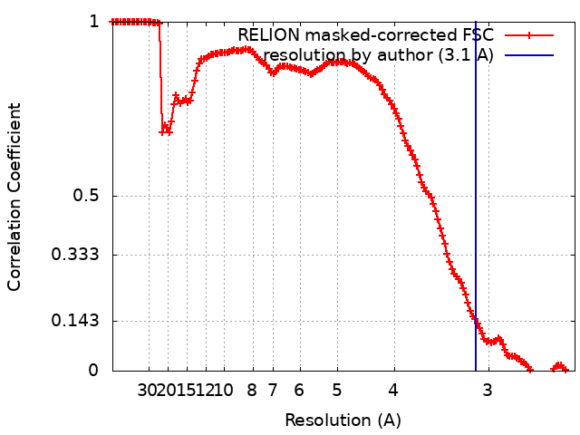
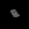
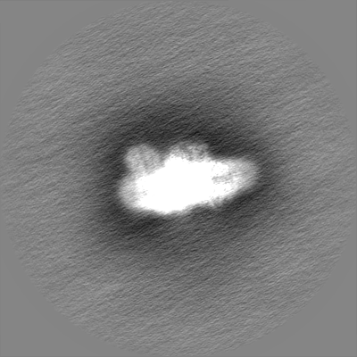
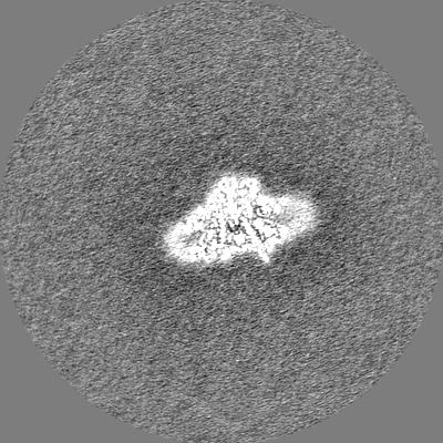
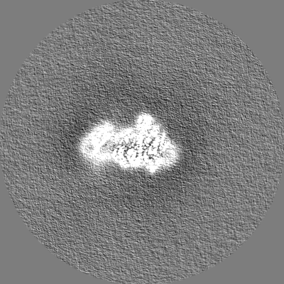
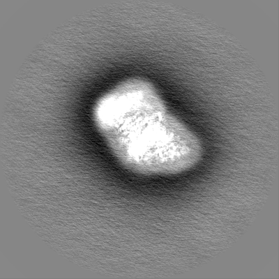
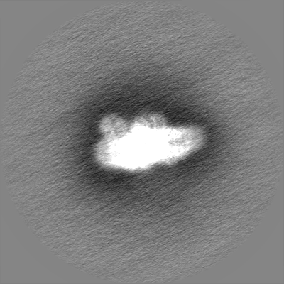
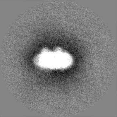
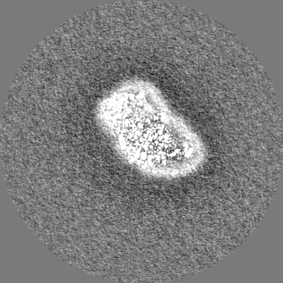
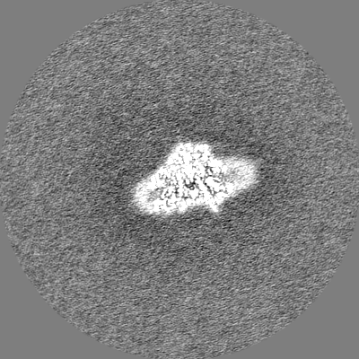
single particle reconstruction / cryo EM / Resolution: 3.1 Å
United Kingdom, European Union, Czech Republic, 5 items
Organization
Grant number
Country
Biotechnology and Biological Sciences Research Council (BBSRC)
BB/L003260/1
United Kingdom
Biotechnology and Biological Sciences Research Council (BBSRC)
BB/P00931X/1
United Kingdom
Biotechnology and Biological Sciences Research Council (BBSRC)
BB/P00931X/1
United Kingdom
European Research Council (ERC)
854126
European Union
Czech Science Foundation
19-29225X
Czech Republic
Citation
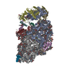
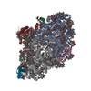
Journal: Nat Commun / Year: 2023 Title: The Ycf48 accessory factor occupies the site of the oxygen-evolving manganese cluster during photosystem II biogenesis. Authors: Ziyu Zhao / Irene Vercellino / Jana Knoppová / Roman Sobotka / James W Murray / Peter J Nixon / Leonid A Sazanov / Josef Komenda / Abstract: Robust oxygenic photosynthesis requires a suite of accessory factors to ensure efficient assembly and repair of the oxygen-evolving photosystem two (PSII) complex. The highly conserved Ycf48 assembly ...Robust oxygenic photosynthesis requires a suite of accessory factors to ensure efficient assembly and repair of the oxygen-evolving photosystem two (PSII) complex. The highly conserved Ycf48 assembly factor binds to the newly synthesized D1 reaction center polypeptide and promotes the initial steps of PSII assembly, but its binding site is unclear. Here we use cryo-electron microscopy to determine the structure of a cyanobacterial PSII D1/D2 reaction center assembly complex with Ycf48 attached. Ycf48, a 7-bladed beta propeller, binds to the amino-acid residues of D1 that ultimately ligate the water-oxidising MnCaO cluster, thereby preventing the premature binding of Mn and Ca ions and protecting the site from damage. Interactions with D2 help explain how Ycf48 promotes assembly of the D1/D2 complex. Overall, our work provides valuable insights into the early stages of PSII assembly and the structural changes that create the binding site for the MnCaO cluster.
Model: Quantifoil R0.6/1 / Material: COPPER / Mesh: 300 / Support film - Material: CARBON / Support film - topology: CONTINUOUS / Support film - Film thickness: 1 / Pretreatment - Type: GLOW DISCHARGE / Pretreatment - Time: 5 sec.
Vitrification
Cryogen name: ETHANE / Chamber humidity: 100 % / Chamber temperature: 277 K / Instrument: FEI VITROBOT MARK IV
-
Electron microscopy
Microscope
TFS GLACIOS
Image recording
Film or detector model: FEI FALCON III (4k x 4k) / Detector mode: INTEGRATING / Number grids imaged: 1 / Number real images: 2853 / Average electron dose: 90.9 e/Å2
Electron beam
Acceleration voltage: 200 kV / Electron source: FIELD EMISSION GUN
Details: 6wj6 was used for RCII, 5ojp for Ycf48 , 5oy0 for PSI
Final reconstruction
Number classes used: 3 / Applied symmetry - Point group: C1 (asymmetric) / Algorithm: FOURIER SPACE / Resolution.type: BY AUTHOR / Resolution: 3.1 Å / Resolution method: FSC 0.143 CUT-OFF / Software - Name: RELION (ver. 3) / Number images used: 178513
Initial angle assignment
Type: MAXIMUM LIKELIHOOD / Software - Name: RELION (ver. 3)
Final angle assignment
Type: MAXIMUM LIKELIHOOD / Software - Name: RELION (ver. 3)
Final 3D classification
Number classes: 3 / Avg.num./class: 80000 / Software - Name: RELION (ver. 3) Details: 80000 particles on average were in the two good classes, while the residual junk only contained c.a. 15000
In the structure databanks used in Yorodumi, some data are registered as the other names, "COVID-19 virus" and "2019-nCoV". Here are the details of the virus and the list of structure data.
Jan 31, 2019. EMDB accession codes are about to change! (news from PDBe EMDB page)
EMDB accession codes are about to change! (news from PDBe EMDB page)
The allocation of 4 digits for EMDB accession codes will soon come to an end. Whilst these codes will remain in use, new EMDB accession codes will include an additional digit and will expand incrementally as the available range of codes is exhausted. The current 4-digit format prefixed with “EMD-” (i.e. EMD-XXXX) will advance to a 5-digit format (i.e. EMD-XXXXX), and so on. It is currently estimated that the 4-digit codes will be depleted around Spring 2019, at which point the 5-digit format will come into force.
The EM Navigator/Yorodumi systems omit the EMD- prefix.
Related info.:Q: What is EMD? / ID/Accession-code notation in Yorodumi/EM Navigator
Yorodumi is a browser for structure data from EMDB, PDB, SASBDB, etc.
This page is also the successor to EM Navigator detail page, and also detail information page/front-end page for Omokage search.
The word "yorodu" (or yorozu) is an old Japanese word meaning "ten thousand". "mi" (miru) is to see.
Related info.:EMDB / PDB / SASBDB / Comparison of 3 databanks / Yorodumi Search / Aug 31, 2016. New EM Navigator & Yorodumi / Yorodumi Papers / Jmol/JSmol / Function and homology information / Changes in new EM Navigator and Yorodumi
 Movie
Movie Controller


 Open data
 Basic information
 Map data
 Sample
 Keywords
 Function and homology information
 Authors
 United Kingdom, European Union,
  Czech Republic, 5 items
  Citation

 Structure visualization
 Downloads & links
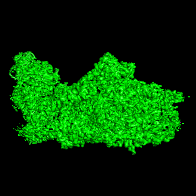 emd_15522.png
 http://ftp.pdbj.org/pub/emdb/structures/EMD-15522


 Z (Sec.)
 Y (Row.)
 X (Col.)


 Sample components


 Processing
 Electron microscopy
 FIELD EMISSION GUN