- EMDB-15484: Structure of human DDB1-DCAF12 in complex with the C-terminus of CCT5 -
+
Open data
ID or keywords:
Loading...
-
Basic information
Entry
Database: EMDB / ID: EMD-15484
Title
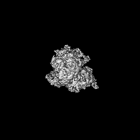
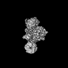
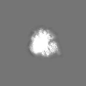
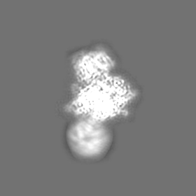
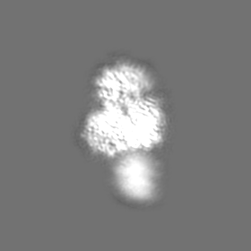
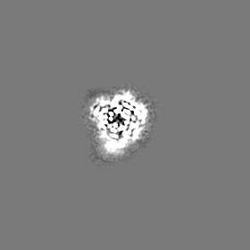
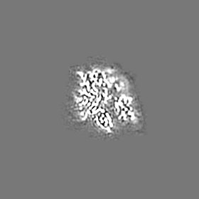
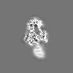
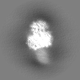
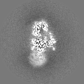
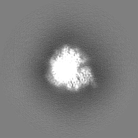
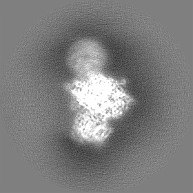
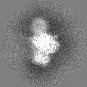
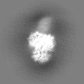
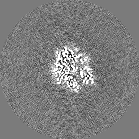
Structure of human DDB1-DCAF12 in complex with the C-terminus of CCT5
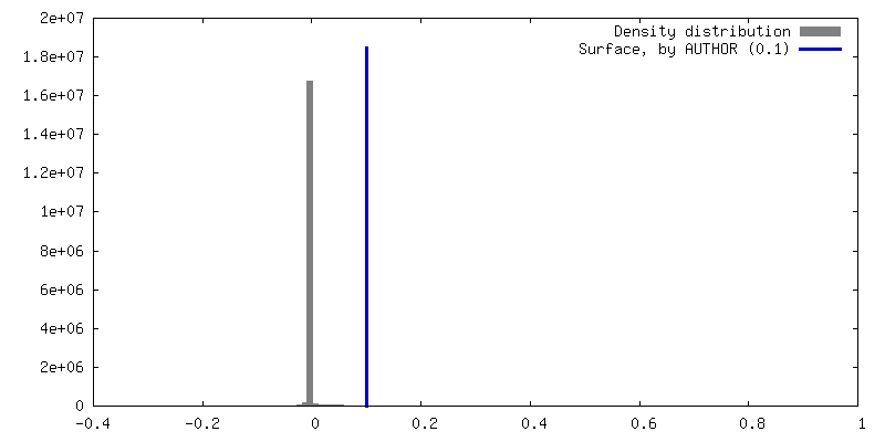
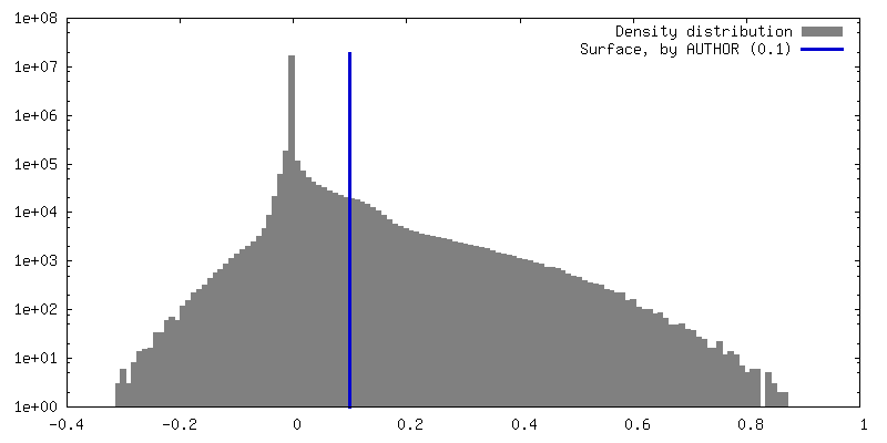
Map data
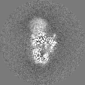
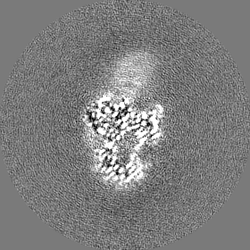
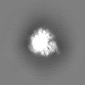
Locally sharpened map (LocScale).
Sample
Complex: Complex of DDB1-DCAF12-CCT5
Protein or peptide: DNA damage-binding protein 1
Protein or peptide: DDB1- and CUL4-associated factor 12
Protein or peptide: T-complex protein 1 subunit epsilon
Keywords
ubiquitin / E3 / TRiC / chaperonin / LIGASE
Function / homology
Function and homology information
positive regulation of protein localization to Cajal body / positive regulation of telomerase RNA localization to Cajal body / chaperonin-containing T-complex / BBSome-mediated cargo-targeting to cilium / Formation of tubulin folding intermediates by CCT/TriC / binding of sperm to zona pellucida / Folding of actin by CCT/TriC / positive regulation by virus of viral protein levels in host cell / Prefoldin mediated transfer of substrate to CCT/TriC / spindle assembly involved in female meiosis ...positive regulation of protein localization to Cajal body / positive regulation of telomerase RNA localization to Cajal body / chaperonin-containing T-complex / BBSome-mediated cargo-targeting to cilium / Formation of tubulin folding intermediates by CCT/TriC / binding of sperm to zona pellucida / Folding of actin by CCT/TriC / positive regulation by virus of viral protein levels in host cell / Prefoldin mediated transfer of substrate to CCT/TriC / spindle assembly involved in female meiosis / epigenetic programming in the zygotic pronuclei / UV-damage excision repair / ubiquitin-dependent protein catabolic process via the C-end degron rule pathway / biological process involved in interaction with symbiont / regulation of mitotic cell cycle phase transition / WD40-repeat domain binding / Cul4A-RING E3 ubiquitin ligase complex / Cul4-RING E3 ubiquitin ligase complex / Cul4B-RING E3 ubiquitin ligase complex / Association of TriC/CCT with target proteins during biosynthesis / ubiquitin ligase complex scaffold activity / negative regulation of reproductive process / negative regulation of developmental process / viral release from host cell / cullin family protein binding / ectopic germ cell programmed cell death / positive regulation of viral genome replication / Hydrolases; Acting on acid anhydrides; In phosphorus-containing anhydrides / beta-tubulin binding / ubiquitin-like ligase-substrate adaptor activity / positive regulation of telomere maintenance via telomerase / proteasomal protein catabolic process / protein folding chaperone / positive regulation of gluconeogenesis / T cell activation / regulation of autophagy / nucleotide-excision repair / mRNA 3'-UTR binding / ATP-dependent protein folding chaperone / sperm end piece / Recognition of DNA damage by PCNA-containing replication complex / regulation of circadian rhythm / DNA Damage Recognition in GG-NER / mRNA 5'-UTR binding / Dual Incision in GG-NER / Transcription-Coupled Nucleotide Excision Repair (TC-NER) / Formation of TC-NER Pre-Incision Complex / Wnt signaling pathway / response to virus / Formation of Incision Complex in GG-NER / Dual incision in TC-NER / Gap-filling DNA repair synthesis and ligation in TC-NER / positive regulation of protein catabolic process / cellular response to UV / : / rhythmic process / Cooperation of PDCL (PhLP1) and TRiC/CCT in G-protein beta folding / G-protein beta-subunit binding / site of double-strand break / sperm principal piece / protein folding / Neddylation / cell body / sperm midpiece / ubiquitin-dependent protein catabolic process / damaged DNA binding / microtubule / proteasome-mediated ubiquitin-dependent protein catabolic process / protein-macromolecule adaptor activity / chromosome, telomeric region / protein stabilization / protein ubiquitination / DNA repair / apoptotic process / DNA damage response / centrosome / negative regulation of apoptotic process / protein-containing complex binding / nucleolus / ATP hydrolysis activity / protein-containing complex / : / DNA binding / extracellular exosome / nucleoplasm / ATP binding / nucleus / cytoplasm / cytosol Similarity search - Function
Journal: EMBO J / Year: 2023 Title: Recognition of the CCT5 di-Glu degron by CRL4 is dependent on TRiC assembly. Authors: Carlos Pla-Prats / Simone Cavadini / Georg Kempf / Nicolas H Thomä / Abstract: Assembly Quality Control (AQC) E3 ubiquitin ligases target incomplete or incorrectly assembled protein complexes for degradation. The CUL4-RBX1-DDB1-DCAF12 (CRL4 ) E3 ligase preferentially ...Assembly Quality Control (AQC) E3 ubiquitin ligases target incomplete or incorrectly assembled protein complexes for degradation. The CUL4-RBX1-DDB1-DCAF12 (CRL4 ) E3 ligase preferentially ubiquitinates proteins that carry a C-terminal double glutamate (di-Glu) motif. Reported CRL4 di-Glu-containing substrates include CCT5, a subunit of the TRiC chaperonin. How DCAF12 engages its substrates and the functional relationship between CRL4 and CCT5/TRiC is currently unknown. Here, we present the cryo-EM structure of the DDB1-DCAF12-CCT5 complex at 2.8 Å resolution. DCAF12 serves as a canonical WD40 DCAF substrate receptor and uses a positively charged pocket at the center of the β-propeller to bind the C-terminus of CCT5. DCAF12 specifically reads out the CCT5 di-Glu side chains, and contacts other visible degron amino acids through Van der Waals interactions. The CCT5 C-terminus is inaccessible in an assembled TRiC complex, and functional assays demonstrate that DCAF12 binds and ubiquitinates monomeric CCT5, but not CCT5 assembled into TRiC. Our biochemical and structural results suggest a previously unknown role for the CRL4 E3 ligase in overseeing the assembly of a key cellular complex.
In the structure databanks used in Yorodumi, some data are registered as the other names, "COVID-19 virus" and "2019-nCoV". Here are the details of the virus and the list of structure data.
Jan 31, 2019. EMDB accession codes are about to change! (news from PDBe EMDB page)
EMDB accession codes are about to change! (news from PDBe EMDB page)
The allocation of 4 digits for EMDB accession codes will soon come to an end. Whilst these codes will remain in use, new EMDB accession codes will include an additional digit and will expand incrementally as the available range of codes is exhausted. The current 4-digit format prefixed with “EMD-” (i.e. EMD-XXXX) will advance to a 5-digit format (i.e. EMD-XXXXX), and so on. It is currently estimated that the 4-digit codes will be depleted around Spring 2019, at which point the 5-digit format will come into force.
The EM Navigator/Yorodumi systems omit the EMD- prefix.
Related info.:Q: What is EMD? / ID/Accession-code notation in Yorodumi/EM Navigator
Yorodumi is a browser for structure data from EMDB, PDB, SASBDB, etc.
This page is also the successor to EM Navigator detail page, and also detail information page/front-end page for Omokage search.
The word "yorodu" (or yorozu) is an old Japanese word meaning "ten thousand". "mi" (miru) is to see.
Related info.:EMDB / PDB / SASBDB / Comparison of 3 databanks / Yorodumi Search / Aug 31, 2016. New EM Navigator & Yorodumi / Yorodumi Papers / Jmol/JSmol / Function and homology information / Changes in new EM Navigator and Yorodumi
 Movie
Movie Controller

 Yorodumi
 Open data
 Basic information
 Map data
 Sample
 Keywords
 Function and homology information
 Homo sapiens (human)
 Authors
 Citation
 Structure visualization
 Downloads & links
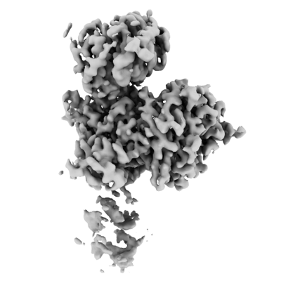 emd_15484.png
 http://ftp.pdbj.org/pub/emdb/structures/EMD-15484


 Z (Sec.)
 Y (Row.)
 X (Col.)


 Sample components
 Trichoplusia ni (cabbage looper)
 Processing
 Electron microscopy
 FIELD EMISSION GUN
