 Movie
Movie Controller


+ Open data

- Basic information
| Entry | Database: EMDB / ID: EMD-1518 | |||||||||
|---|---|---|---|---|---|---|---|---|---|---|
| Title | Cryoelectron tomography of HIV-1 envelope spikes | |||||||||
 Map data Map data | Raw tomogram of HIV-1 BaL virions, no filter applied. | |||||||||
 Sample Sample |
| |||||||||
 Keywords Keywords | HIV-1 / envelope spikes / cryo electron tomography | |||||||||
| Biological species |   Human immunodeficiency virus 1 Human immunodeficiency virus 1 | |||||||||
| Method | electron tomography / cryo EM | |||||||||
 Authors Authors | Zhu P / Winkler H / Chertova E / Taylor KA / Roux KH | |||||||||
 Citation Citation | Journal: PLoS Pathog / Year: 2008 Title: Cryoelectron tomography of HIV-1 envelope spikes: further evidence for tripod-like legs. Authors: Ping Zhu / Hanspeter Winkler / Elena Chertova / Kenneth A Taylor / Kenneth H Roux /  Abstract: A detailed understanding of the morphology of the HIV-1 envelope (Env) spike is key to understanding viral pathogenesis and for informed vaccine design. We have previously presented a cryoelectron ...A detailed understanding of the morphology of the HIV-1 envelope (Env) spike is key to understanding viral pathogenesis and for informed vaccine design. We have previously presented a cryoelectron microscopic tomogram (cryoET) of the Env spikes on SIV virions. Several structural features were noted in the gp120 head and gp41 stalk regions. Perhaps most notable was the presence of three splayed legs projecting obliquely from the base of the spike head toward the viral membrane. Subsequently, a second 3D image of SIV spikes, also obtained by cryoET, was published by another group which featured a compact vertical stalk. We now report the cryoET analysis of HIV-1 virion-associated Env spikes using enhanced analytical cryoET procedures. More than 2,000 Env spike volumes were initially selected, aligned, and sorted into structural classes using algorithms that compensate for the "missing wedge" and do not impose any symmetry. The results show varying morphologies between structural classes: some classes showed trimers in the head domains; nearly all showed two or three legs, though unambiguous three-fold symmetry was not observed either in the heads or the legs. Subsequently, clearer evidence of trimeric head domains and three splayed legs emerged when head and leg volumes were independently aligned and classified. These data show that HIV-1, like SIV, also displays the tripod-like leg configuration, and, unexpectedly, shows considerable gp41 leg flexibility/heteromorphology. The tripod-like model for gp41 is consistent with, and helps explain, many of the unique biophysical and immunological features of this region. | |||||||||
| History |
|
- Structure visualization
| Movie |
 Movie viewer Movie viewer |
|---|---|
| Supplemental images |
- Downloads & links
-EMDB archive
| Map data | emd_1518.map.gz | 143.2 KB | EMDB map data format | |
|---|---|---|---|---|
| Header (meta data) | emd-1518-v30.xmlemd-1518.xml | 9.1 KB 9.1 KB | Display Display | EMDB header |
| Images |  1518.gif 1518.gif | 103.3 KB | ||
| Archive directory |  http://ftp.pdbj.org/pub/emdb/structures/EMD-1518ftp://ftp.pdbj.org/pub/emdb/structures/EMD-1518 http://ftp.pdbj.org/pub/emdb/structures/EMD-1518ftp://ftp.pdbj.org/pub/emdb/structures/EMD-1518 | HTTPS FTP |
-Validation report
| Summary document | emd_1518_validation.pdf.gz | 144.9 KB | Display | EMDB validaton report |
|---|---|---|---|---|
| Full document | emd_1518_full_validation.pdf.gz | 144 KB | Display | |
| Data in XML | emd_1518_validation.xml.gz | 4.8 KB | Display | |
| Arichive directory | https://ftp.pdbj.org/pub/emdb/validation_reports/EMD-1518ftp://ftp.pdbj.org/pub/emdb/validation_reports/EMD-1518 | HTTPS FTP |
-Related structure data
| Related structure data |  1513C  1514C  1515C 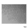 1516C 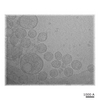 1517C  1519C 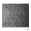 1520C 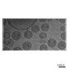 1521C 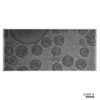 1522C 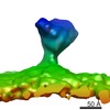 1596C C: citing same article ( |
|---|
-Links
| EMDB pages | EMDB (EBI/PDBe) / EMDataResource |
|---|---|
| Related items in Molecule of the Month |

-Map
| File | Download / File: emd_1518.map.gz / Format: CCP4 / Size: 154.3 KB / Type: IMAGE STORED AS FLOATING POINT NUMBER (4 BYTES) | ||||||||||||||||||||||||||||||||||||||||||||||||||||||||||||||||||||
|---|---|---|---|---|---|---|---|---|---|---|---|---|---|---|---|---|---|---|---|---|---|---|---|---|---|---|---|---|---|---|---|---|---|---|---|---|---|---|---|---|---|---|---|---|---|---|---|---|---|---|---|---|---|---|---|---|---|---|---|---|---|---|---|---|---|---|---|---|---|
| Annotation | Raw tomogram of HIV-1 BaL virions, no filter applied. | ||||||||||||||||||||||||||||||||||||||||||||||||||||||||||||||||||||
| Voxel size | X=Y=Z: 5.56 Å | ||||||||||||||||||||||||||||||||||||||||||||||||||||||||||||||||||||
| Density |
| ||||||||||||||||||||||||||||||||||||||||||||||||||||||||||||||||||||
| Symmetry | Space group: 1 | ||||||||||||||||||||||||||||||||||||||||||||||||||||||||||||||||||||
| Details | EMDB XML:
CCP4 map header:
| ||||||||||||||||||||||||||||||||||||||||||||||||||||||||||||||||||||
-Supplemental data
- Sample components
-Entire : HIV-1 BaL / SUPT1-CCR5 CL.30, provided by the AIDS Vaccine Progra...
| Entire | Name: HIV-1 BaL / SUPT1-CCR5 CL.30, provided by the AIDS Vaccine Program, SAIC Frederick, Inc., NCI, Frederick, MD. Lot p3955. |
|---|---|
| Components |
|
-Supramolecule #1000: HIV-1 BaL / SUPT1-CCR5 CL.30, provided by the AIDS Vaccine Progra...
| Supramolecule | Name: HIV-1 BaL / SUPT1-CCR5 CL.30, provided by the AIDS Vaccine Program, SAIC Frederick, Inc., NCI, Frederick, MD. Lot p3955. type: sample / ID: 1000 Details: The map is the raw tomogram of HIV-1 BaL virus. The envelope spikes (trimer of gp120 and ecto domain of gp41, total MW is about 0.45 MegaDaltons)on the virion surface were picked up and ...Details: The map is the raw tomogram of HIV-1 BaL virus. The envelope spikes (trimer of gp120 and ecto domain of gp41, total MW is about 0.45 MegaDaltons)on the virion surface were picked up and subject to 3D alignment and classification analysis. Oligomeric state: Highly purified virus. The envelope spikes on the virion surface are trimer. Number unique components: 1 |
|---|
-Supramolecule #1: Human immunodeficiency virus 1
| Supramolecule | Name: Human immunodeficiency virus 1 / type: virus / ID: 1 / Name.synonym: HIV-1 Details: The envelope spikes are distributed on the surface of HIV-1 virions. NCBI-ID: 11676 / Sci species name: Human immunodeficiency virus 1 / Database: NCBI / Virus type: VIRION / Virus isolate: STRAIN / Virus enveloped: Yes / Virus empty: No / Syn species name: HIV-1 |
|---|---|
| Host (natural) | Organism:  Homo sapiens (human) / synonym: BACTERIA(EUBACTERIA) Homo sapiens (human) / synonym: BACTERIA(EUBACTERIA) |
-Experimental details
-Structure determination
| Method | cryo EM |
|---|---|
 Processing Processing | electron tomography |
-Sample preparation
| Concentration | 2.8 mg/mL |
|---|---|
| Buffer | pH: 7.4 / Details: Originally in TNE, resuspend in PBS |
| Grid | Details: 300 mesh R2/1 Quantifoil grid |
| Vitrification | Cryogen name: ETHANE / Instrument: HOMEMADE PLUNGER Details: Vitrification instrument: Home made. Vitrification carried out in cold room Method: Blot for 5-6 seconds before plunging |
- Electron microscopy
| Microscope | FEI/PHILIPS CM300FEG/T |
|---|---|
| Temperature | Average: 97 K |
| Image recording | Category: CCD / Film or detector model: TVIPS TEMCAM-F224 (2k x 2k) / Average electron dose: 1.5 e/Å2 |
| Electron beam | Acceleration voltage: 300 kV / Electron source:  FIELD EMISSION GUN FIELD EMISSION GUN |
| Electron optics | Illumination mode: FLOOD BEAM / Imaging mode: BRIGHT FIELD / Cs: 2 mm / Nominal defocus max: 4.0 µm / Nominal magnification: 43200 |
| Sample stage | Specimen holder: Eucentric / Specimen holder model: GATAN LIQUID NITROGEN / Tilt series - Axis1 - Min angle: -70 ° / Tilt series - Axis1 - Max angle: 57 ° / Tilt series - Axis1 - Angle increment: 2 ° |
-Image processing
| Details | The tilt angles were determined using saxton schedule. The angle increment is 2 deg at low tilt angles range and decreased to about 1 deg at high tilt angles range. |
|---|---|
| Final reconstruction | Algorithm: OTHER / Software - Name: PROTOMO / Number images used: 84 |
