 Movie
Movie Controller

[English] 日本語
 Yorodumi
- EMDB-15150: Cryo-electron tomogram of secreted particles from Apilactobacillu... -
+ Open data

- Basic information
| Entry | 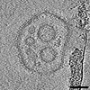 | |||||||||
|---|---|---|---|---|---|---|---|---|---|---|
| Title | Cryo-electron tomogram of secreted particles from Apilactobacillus kunkeei (TS_01) | |||||||||
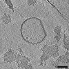
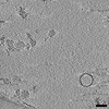
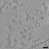
 Map data Map data | Cryo-ET tomogram of secreted particles from Apilactobacillus kunkeei isolated from cell-free pellet preparations. Both membrane vesicles and speckled, contrast-rich particles were observed. | |||||||||
 Sample Sample |
| |||||||||
 Keywords Keywords | Membrane vesicles / extracellular protein complexes / UNKNOWN FUNCTION | |||||||||
| Biological species |  Apilactobacillus kunkeei (bacteria) Apilactobacillus kunkeei (bacteria) | |||||||||
| Method | electron tomography / cryo EM / Resolution: 10.0923 Å | |||||||||
 Authors Authors | Seeger C / Andersson SGE | |||||||||
| Funding support |  Sweden, 1 items Sweden, 1 items
| |||||||||
 Citation Citation | Journal: To Be Published Title: Cryo-eletron tomogram of secreted particles from Apilactobacillus kunkeei (TS_01) Authors: Seeger C / Andersson SGE | |||||||||
| History |
|
- Structure visualization
| Supplemental images |
|---|

- Downloads & links
-EMDB archive
| Map data | emd_15150.map.gz | 10.5 MB |  EMDB map data format EMDB map data format | |
|---|---|---|---|---|
| Header (meta data) | emd-15150-v30.xmlemd-15150.xml | 9 KB 9 KB | Display Display | EMDB header |
| Images |  emd_15150.png emd_15150.png | 187.4 KB | ||
| Archive directory |  http://ftp.pdbj.org/pub/emdb/structures/EMD-15150ftp://ftp.pdbj.org/pub/emdb/structures/EMD-15150 http://ftp.pdbj.org/pub/emdb/structures/EMD-15150ftp://ftp.pdbj.org/pub/emdb/structures/EMD-15150 | HTTPS FTP |
-Validation report
| Summary document | emd_15150_validation.pdf.gz | 503.3 KB | Display | EMDB validaton report |
|---|---|---|---|---|
| Full document | emd_15150_full_validation.pdf.gz | 502.8 KB | Display | |
| Data in XML | emd_15150_validation.xml.gz | 2.4 KB | Display | |
| Data in CIF | emd_15150_validation.cif.gz | 2.8 KB | Display | |
| Arichive directory | https://ftp.pdbj.org/pub/emdb/validation_reports/EMD-15150ftp://ftp.pdbj.org/pub/emdb/validation_reports/EMD-15150 | HTTPS FTP |
-Related structure data


-Links
| EMDB pages | EMDB (EBI/PDBe) / EMDataResource |
|---|
-Map
| File | Download / File: emd_15150.map.gz / Format: CCP4 / Size: 25 MB / Type: IMAGE STORED AS SIGNED BYTE | ||||||||||||||||||||
|---|---|---|---|---|---|---|---|---|---|---|---|---|---|---|---|---|---|---|---|---|---|
| Annotation | Cryo-ET tomogram of secreted particles from Apilactobacillus kunkeei isolated from cell-free pellet preparations. Both membrane vesicles and speckled, contrast-rich particles were observed. | ||||||||||||||||||||
| Voxel size | X=Y=Z: 10.09232 Å | ||||||||||||||||||||
| Density |
| ||||||||||||||||||||
| Symmetry | Space group: 1 | ||||||||||||||||||||
| Details | EMDB XML:
|
-Supplemental data
- Sample components
-Entire : Secreted nanoparticles (membrane vesicles, protein complexes) fro...
| Entire | Name: Secreted nanoparticles (membrane vesicles, protein complexes) from Apilactobacillus kunkeei |
|---|---|
| Components |
|
-Supramolecule #1: Secreted nanoparticles (membrane vesicles, protein complexes) fro...
| Supramolecule | Name: Secreted nanoparticles (membrane vesicles, protein complexes) from Apilactobacillus kunkeei type: complex / ID: 1 / Parent: 0 |
|---|---|
| Source (natural) | Organism: Apilactobacillus kunkeei (bacteria) / Strain: A1401 |
-Experimental details
-Structure determination
| Method | cryo EM |
|---|---|
 Processing Processing | electron tomography |
| Aggregation state | particle |
-Sample preparation
| Buffer | pH: 7.4 / Component - Concentration: 10.0 mM / Component - Name: Phosphate buffer / Details: 10 mM phosphate buffer, pH 7.4 |
|---|---|
| Grid | Model: Quantifoil / Material: COPPER / Mesh: 200 / Support film - Material: CARBON / Support film - topology: HOLEY / Support film - Film thickness: 2 / Pretreatment - Type: GLOW DISCHARGE / Pretreatment - Time: 60 sec. / Pretreatment - Atmosphere: AIR / Pretreatment - Pressure: 101.325 kPa |
| Vitrification | Cryogen name: ETHANE / Chamber humidity: 100 % / Chamber temperature: 277 K / Instrument: FEI VITROBOT MARK IV / Details: blot time 5s. |
| Sectioning | Other: NO SECTIONING |
| Fiducial marker | Manufacturer: Aurion / Diameter: 10 nm |
- Electron microscopy
| Microscope | FEI TALOS ARCTICA |
|---|---|
| Image recording | Film or detector model: FEI FALCON III (4k x 4k) / Average exposure time: 3.0 sec. / Average electron dose: 95.0 e/Å2 |
| Electron beam | Acceleration voltage: 200 kV / Electron source: OTHER |
| Electron optics | C2 aperture diameter: 50.0 µm / Illumination mode: OTHER / Imaging mode: OTHER / Nominal defocus max: 6.0 µm / Nominal defocus min: 4.0 µm |
| Sample stage | Cooling holder cryogen: NITROGEN |
| Experimental equipment |  Model: Talos Arctica / Image courtesy: FEI Company |
-Image processing
| Details | The initial raw movies were aligned and dose-weight filtered using alignframes from the IMOD package |
|---|---|
| Final reconstruction | Algorithm: BACK PROJECTION / Resolution.type: BY AUTHOR / Resolution: 10.0923 Å / Resolution method: OTHER / Software - Name:  IMOD (ver. v4.11.6) / Number images used: 61 IMOD (ver. v4.11.6) / Number images used: 61 |
