[English] 日本語
 Yorodumi
- EMDB-14867: Dimeric PSI of Chlamydomonas reinhardtii at 2.74 A resolution (sy... -
+ Open data
- Basic information
| Entry |  | |||||||||||||||
|---|---|---|---|---|---|---|---|---|---|---|---|---|---|---|---|---|
| Title | Dimeric PSI of Chlamydomonas reinhardtii at 2.74 A resolution (symmetry expanded) | |||||||||||||||
 Map data Map data | ||||||||||||||||
 Sample Sample |
| |||||||||||||||
| Function / homology |  Function and homology information Function and homology informationchloroplast thylakoid lumen / photosynthesis, light harvesting / photosynthesis, light harvesting in photosystem I / photosystem I reaction center / photosystem I / photosynthetic electron transport in photosystem I / photosystem I / photosystem II / chloroplast thylakoid membrane / chlorophyll binding ...chloroplast thylakoid lumen / photosynthesis, light harvesting / photosynthesis, light harvesting in photosystem I / photosystem I reaction center / photosystem I / photosynthetic electron transport in photosystem I / photosystem I / photosystem II / chloroplast thylakoid membrane / chlorophyll binding / response to light stimulus / photosynthesis / 4 iron, 4 sulfur cluster binding / electron transfer activity / oxidoreductase activity / magnesium ion binding / metal ion binding Similarity search - Function | |||||||||||||||
| Biological species |  | |||||||||||||||
| Method | single particle reconstruction / cryo EM / Resolution: 2.74 Å | |||||||||||||||
 Authors Authors | Naschberger A / Amunts A | |||||||||||||||
| Funding support |  Sweden, Sweden,  Germany, 4 items Germany, 4 items
| |||||||||||||||
 Citation Citation |  Journal: Nat Plants / Year: 2022 Journal: Nat Plants / Year: 2022Title: Algal photosystem I dimer and high-resolution model of PSI-plastocyanin complex. Authors: Andreas Naschberger / Laura Mosebach / Victor Tobiasson / Sebastian Kuhlgert / Martin Scholz / Annemarie Perez-Boerema / Thi Thu Hoai Ho / André Vidal-Meireles / Yuichiro Takahashi / ...Authors: Andreas Naschberger / Laura Mosebach / Victor Tobiasson / Sebastian Kuhlgert / Martin Scholz / Annemarie Perez-Boerema / Thi Thu Hoai Ho / André Vidal-Meireles / Yuichiro Takahashi / Michael Hippler / Alexey Amunts /     Abstract: Photosystem I (PSI) enables photo-electron transfer and regulates photosynthesis in the bioenergetic membranes of cyanobacteria and chloroplasts. Being a multi-subunit complex, its macromolecular ...Photosystem I (PSI) enables photo-electron transfer and regulates photosynthesis in the bioenergetic membranes of cyanobacteria and chloroplasts. Being a multi-subunit complex, its macromolecular organization affects the dynamics of photosynthetic membranes. Here we reveal a chloroplast PSI from the green alga Chlamydomonas reinhardtii that is organized as a homodimer, comprising 40 protein subunits with 118 transmembrane helices that provide scaffold for 568 pigments. Cryogenic electron microscopy identified that the absence of PsaH and Lhca2 gives rise to a head-to-head relative orientation of the PSI-light-harvesting complex I monomers in a way that is essentially different from the oligomer formation in cyanobacteria. The light-harvesting protein Lhca9 is the key element for mediating this dimerization. The interface between the monomers is lacking PsaH and thus partially overlaps with the surface area that would bind one of the light-harvesting complex II complexes in state transitions. We also define the most accurate available PSI-light-harvesting complex I model at 2.3 Å resolution, including a flexibly bound electron donor plastocyanin, and assign correct identities and orientations to all the pigments, as well as 621 water molecules that affect energy transfer pathways. | |||||||||||||||
| History |
|
- Structure visualization
| Supplemental images |
|---|
- Downloads & links
-EMDB archive
| Map data |  emd_14867.map.gz emd_14867.map.gz | 694 MB |  EMDB map data format EMDB map data format | |
|---|---|---|---|---|
| Header (meta data) |  emd-14867-v30.xml emd-14867-v30.xml emd-14867.xml emd-14867.xml | 39 KB 39 KB | Display Display |  EMDB header EMDB header |
| FSC (resolution estimation) |  emd_14867_fsc.xml emd_14867_fsc.xml | 24.6 KB | Display |  FSC data file FSC data file |
| Images |  emd_14867.png emd_14867.png | 87.2 KB | ||
| Masks |  emd_14867_msk_1.map emd_14867_msk_1.map | 1.3 GB |  Mask map Mask map | |
| Others |  emd_14867_half_map_1.map.gz emd_14867_half_map_1.map.gz emd_14867_half_map_2.map.gz emd_14867_half_map_2.map.gz | 1 GB 1 GB | ||
| Archive directory |  http://ftp.pdbj.org/pub/emdb/structures/EMD-14867 http://ftp.pdbj.org/pub/emdb/structures/EMD-14867 ftp://ftp.pdbj.org/pub/emdb/structures/EMD-14867 ftp://ftp.pdbj.org/pub/emdb/structures/EMD-14867 | HTTPS FTP |
-Validation report
| Summary document |  emd_14867_validation.pdf.gz emd_14867_validation.pdf.gz | 811.3 KB | Display |  EMDB validaton report EMDB validaton report |
|---|---|---|---|---|
| Full document |  emd_14867_full_validation.pdf.gz emd_14867_full_validation.pdf.gz | 810.8 KB | Display | |
| Data in XML |  emd_14867_validation.xml.gz emd_14867_validation.xml.gz | 32.9 KB | Display | |
| Data in CIF |  emd_14867_validation.cif.gz emd_14867_validation.cif.gz | 43.9 KB | Display | |
| Arichive directory |  https://ftp.pdbj.org/pub/emdb/validation_reports/EMD-14867 https://ftp.pdbj.org/pub/emdb/validation_reports/EMD-14867 ftp://ftp.pdbj.org/pub/emdb/validation_reports/EMD-14867 ftp://ftp.pdbj.org/pub/emdb/validation_reports/EMD-14867 | HTTPS FTP |
-Related structure data
| Related structure data | 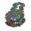 7zq9MC 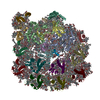 7zqcC 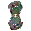 7zqdC  7zqeC M: atomic model generated by this map C: citing same article ( |
|---|---|
| Similar structure data | Similarity search - Function & homology  F&H Search F&H Search |
- Links
Links
| EMDB pages |  EMDB (EBI/PDBe) / EMDB (EBI/PDBe) /  EMDataResource EMDataResource |
|---|---|
| Related items in Molecule of the Month |
- Map
Map
| File |  Download / File: emd_14867.map.gz / Format: CCP4 / Size: 1.3 GB / Type: IMAGE STORED AS FLOATING POINT NUMBER (4 BYTES) Download / File: emd_14867.map.gz / Format: CCP4 / Size: 1.3 GB / Type: IMAGE STORED AS FLOATING POINT NUMBER (4 BYTES) | ||||||||||||||||||||||||||||||||||||
|---|---|---|---|---|---|---|---|---|---|---|---|---|---|---|---|---|---|---|---|---|---|---|---|---|---|---|---|---|---|---|---|---|---|---|---|---|---|
| Projections & slices | Image control
Images are generated by Spider. | ||||||||||||||||||||||||||||||||||||
| Voxel size | X=Y=Z: 0.84 Å | ||||||||||||||||||||||||||||||||||||
| Density |
| ||||||||||||||||||||||||||||||||||||
| Symmetry | Space group: 1 | ||||||||||||||||||||||||||||||||||||
| Details | EMDB XML:
|
-Supplemental data
-Mask #1
| File |  emd_14867_msk_1.map emd_14867_msk_1.map | ||||||||||||
|---|---|---|---|---|---|---|---|---|---|---|---|---|---|
| Projections & Slices |
| ||||||||||||
| Density Histograms |
-Half map: #2
| File | emd_14867_half_map_1.map | ||||||||||||
|---|---|---|---|---|---|---|---|---|---|---|---|---|---|
| Projections & Slices |
| ||||||||||||
| Density Histograms |
-Half map: #1
| File | emd_14867_half_map_2.map | ||||||||||||
|---|---|---|---|---|---|---|---|---|---|---|---|---|---|
| Projections & Slices |
| ||||||||||||
| Density Histograms |
- Sample components
+Entire : Photosystem I dimer of Chlamydomonas reinhardtii
+Supramolecule #1: Photosystem I dimer of Chlamydomonas reinhardtii
+Macromolecule #1: Photosystem I P700 chlorophyll a apoprotein A1
+Macromolecule #2: Photosystem I P700 chlorophyll a apoprotein A2
+Macromolecule #3: Photosystem I iron-sulfur center
+Macromolecule #4: Photosystem I reaction center subunit II, chloroplastic
+Macromolecule #5: Photosystem I reaction center subunit IV, chloroplastic
+Macromolecule #6: Photosystem I reaction center subunit III, chloroplastic
+Macromolecule #7: Photosystem I reaction center subunit V, chloroplastic
+Macromolecule #8: Photosystem I reaction center subunit VIII
+Macromolecule #9: Photosystem I reaction center subunit IX
+Macromolecule #10: PSI subunit V
+Macromolecule #11: Photosystem I reaction center subunit psaK, chloroplastic
+Macromolecule #12: Chlorophyll a-b binding protein, chloroplastic
+Macromolecule #13: Chlorophyll a-b binding protein, chloroplastic
+Macromolecule #14: Chlorophyll a-b binding protein, chloroplastic
+Macromolecule #15: Chlorophyll a-b binding protein, chloroplastic
+Macromolecule #16: Chlorophyll a-b binding protein, chloroplastic (Lhca4)
+Macromolecule #17: Chlorophyll a-b binding protein, chloroplastic
+Macromolecule #18: Chlorophyll a-b binding protein, chloroplastic
+Macromolecule #19: Chlorophyll a-b binding protein, chloroplastic
+Macromolecule #20: Photosystem I P700 chlorophyll a apoprotein A2
+Macromolecule #21: CHLOROPHYLL A ISOMER
+Macromolecule #22: CHLOROPHYLL A
+Macromolecule #23: PHYLLOQUINONE
+Macromolecule #24: 1,2-DIPALMITOYL-PHOSPHATIDYL-GLYCEROLE
+Macromolecule #25: BETA-CAROTENE
+Macromolecule #26: IRON/SULFUR CLUSTER
+Macromolecule #27: DODECYL-ALPHA-D-MALTOSIDE
+Macromolecule #28: 1,2-DISTEAROYL-MONOGALACTOSYL-DIGLYCERIDE
+Macromolecule #29: (3R,3'R,6S)-4,5-DIDEHYDRO-5,6-DIHYDRO-BETA,BETA-CAROTENE-3,3'-DIOL
+Macromolecule #30: DIGALACTOSYL DIACYL GLYCEROL (DGDG)
+Macromolecule #31: CHLOROPHYLL B
+Macromolecule #32: (3S,5R,6S,3'S,5'R,6'S)-5,6,5',6'-DIEPOXY-5,6,5',6'- TETRAHYDRO-BE...
+Macromolecule #33: (1R,3R)-6-{(3E,5E,7E,9E,11E,13E,15E,17E)-18-[(1S,4R,6R)-4-HYDROXY...
+Macromolecule #34: water
-Experimental details
-Structure determination
| Method | cryo EM |
|---|---|
 Processing Processing | single particle reconstruction |
| Aggregation state | particle |
- Sample preparation
| Buffer | pH: 7.5 |
|---|---|
| Vitrification | Cryogen name: ETHANE |
- Electron microscopy
| Microscope | FEI TITAN KRIOS |
|---|---|
| Image recording | Film or detector model: GATAN K3 BIOQUANTUM (6k x 4k) / Average electron dose: 45.8 e/Å2 |
| Electron beam | Acceleration voltage: 300 kV / Electron source:  FIELD EMISSION GUN FIELD EMISSION GUN |
| Electron optics | Illumination mode: FLOOD BEAM / Imaging mode: BRIGHT FIELD / Nominal defocus max: 5.0 µm / Nominal defocus min: 1.0 µm |
| Experimental equipment |  Model: Titan Krios / Image courtesy: FEI Company |
 Movie
Movie Controller


 Z (Sec.)
 Y (Row.)
 X (Col.)


