ムービー
ムービー コントローラー


+ データを開く

- 基本情報
基本情報
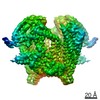
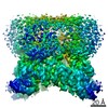
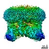
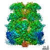
| 登録情報 | データベース: EMDB / ID: EMD-11156 | |||||||||
|---|---|---|---|---|---|---|---|---|---|---|
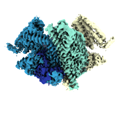
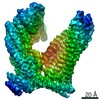
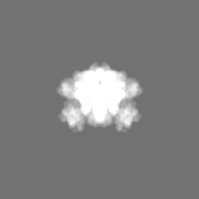
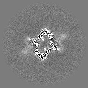
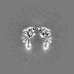
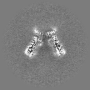
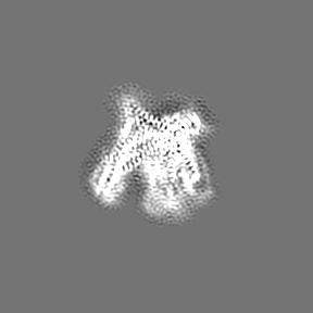
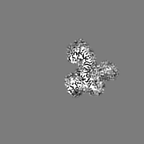
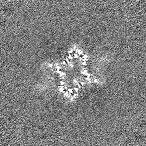
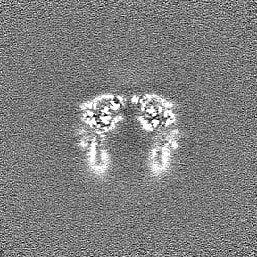
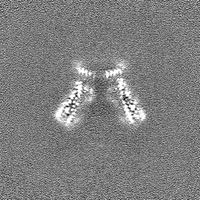
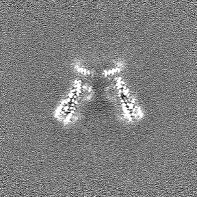
| タイトル | Plasmodium falciparum merozoite surface protein 1 dimer, conformation 1 | |||||||||
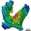
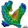
 マップデータ マップデータ | Dimer, C2 reconstruction, unfiltered | |||||||||
 試料 試料 |
| |||||||||
 キーワード キーワード | Merozoite surface protein 1 / malaria / Plasmodium falciparum / MSP-1 / p190 / GPI-anchored membrane protein / MEMBRANE PROTEIN | |||||||||
| 機能・相同性 |  機能・相同性情報 機能・相同性情報 | |||||||||
| 生物種 |  | |||||||||
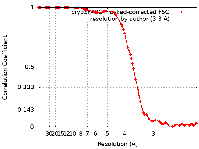
| 手法 | 単粒子再構成法 / クライオ電子顕微鏡法 / 解像度: 3.3 Å | |||||||||
 データ登録者 データ登録者 | Dijkman PM / Kudryashev M | |||||||||
| 資金援助 |  ドイツ, 1件 ドイツ, 1件
| |||||||||
 引用 引用 | ジャーナル: Sci Adv / 年: 2021 タイトル: Structure of the merozoite surface protein 1 from . 著者: Patricia M Dijkman / Tanja Marzluf / Yingyi Zhang / Shih-Ying Scott Chang / Dominic Helm / Michael Lanzer / Hermann Bujard / Mikhail Kudryashev / 要旨: The merozoite surface protein 1 (MSP-1) is the most abundant protein on the surface of the erythrocyte-invading merozoite, the causative agent of malaria. MSP-1 is essential for merozoite formation, ...The merozoite surface protein 1 (MSP-1) is the most abundant protein on the surface of the erythrocyte-invading merozoite, the causative agent of malaria. MSP-1 is essential for merozoite formation, entry into and escape from erythrocytes, and is a promising vaccine candidate. Here, we present monomeric and dimeric structures of full-length MSP-1. MSP-1 adopts an unusual fold with a large central cavity. Its fold includes several coiled-coils and shows structural homology to proteins associated with membrane and cytoskeleton interactions. MSP-1 formed dimers through these domains in a concentration-dependent manner. Dimerization is affected by the presence of the erythrocyte cytoskeleton protein spectrin, which may compete for the dimerization interface. Our work provides structural insights into the possible mode of interaction of MSP-1 with erythrocytes and establishes a framework for future investigations into the role of MSP-1 in infection and immunity. #1: ジャーナル: Sci Adv / 年: 2021タイトル: Structure of the merozoite surface protein 1 from Plasmodium falciparum 著者: Dijkman PM / Marzluf T / Zhang Y / Chang SYS / Helm D / Lanzer M / Bujard H / Kudryashev M | |||||||||
| 履歴 |
|
- 構造の表示
構造の表示
| ムービー |
ムービービューア |
|---|---|
| 構造ビューア | EMマップ: SurfViewMolmilJmol/JSmol |
| 添付画像 |
- ダウンロードとリンク
-EMDBアーカイブ
| マップデータ | emd_11156.map.gz | 45.5 MB | EMDBマップデータ形式 | |
|---|---|---|---|---|
| ヘッダ (付随情報) | emd-11156-v30.xmlemd-11156.xml | 30.7 KB 30.7 KB | 表示 表示 | EMDBヘッダ |
| FSC (解像度算出) | emd_11156_fsc.xml | 10.9 KB | 表示 | FSCデータファイル |
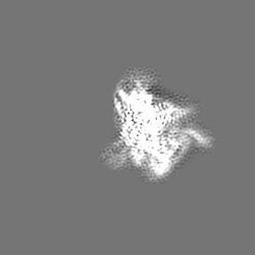
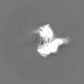
| 画像 |  emd_11156.png emd_11156.png | 132.3 KB | ||
| マスクデータ | emd_11156_msk_1.map | 91.1 MB | マスクマップ | |
| Filedesc metadata | emd-11156.cif.gz | 6.9 KB | ||
| その他 | emd_11156_additional_1.map.gzemd_11156_additional_2.map.gzemd_11156_additional_3.map.gzemd_11156_additional_4.map.gzemd_11156_additional_5.map.gzemd_11156_half_map_1.map.gzemd_11156_half_map_2.map.gz | 86 MB 71 MB 6.8 MB 71.1 MB 71.3 MB 84.5 MB 84.5 MB | ||
| アーカイブディレクトリ |  http://ftp.pdbj.org/pub/emdb/structures/EMD-11156ftp://ftp.pdbj.org/pub/emdb/structures/EMD-11156 http://ftp.pdbj.org/pub/emdb/structures/EMD-11156ftp://ftp.pdbj.org/pub/emdb/structures/EMD-11156 | HTTPS FTP |
-関連構造データ
| 関連構造データ |  6zbjMC  6zbcC 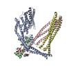 6zbdC 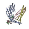 6zbeC 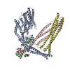 6zbfC 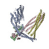 6zbgC 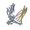 6zbhC 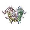 6zblC C: 同じ文献を引用 ( M: このマップから作成された原子モデル |
|---|---|
| 類似構造データ | |
| 電子顕微鏡画像生データ | EMPIAR-10437 (タイトル: Single-particle cryo-EM of the full-length merozoite surface protein 1 from Plasmodium falciparum Data size: 34.0 TB Data #1: Unaligned movies of MSP-1 [micrographs - multiframe] Data #2: Unaligned movies of hdMSP-1 (dataset 1) [micrographs - multiframe] Data #3: Unaligned movies of hdMSP-1 (dataset 2) [micrographs - multiframe] Data #4: Unaligned movies of hdMSP-1 (dataset 3) [micrographs - multiframe] Data #5: Unaligned movies of hdMSP-1 (dataset 4) [micrographs - multiframe] Data #6: Final particle stack for hdMSP-1 conformation 1 [picked particles - single frame - processed] Data #7: Final particle stack for hdMSP-1 conformation 2 [picked particles - single frame - processed] Data #8: Final particle stack for MSP-1 main conformation [picked particles - single frame - processed] Data #9: Final particle stack for MSP-1 alternative conformation 1 [picked particles - single frame - processed] Data #10: Final particle stack for MSP-1 alternative conformation 2 [picked particles - single frame - processed] Data #11: Final particle stack for MSP-1 alternative conformation 3 [picked particles - single frame - processed] Data #12: Final particle stack for MSP-1 alternative conformation 4 [picked particles - single frame - processed] Data #13: Final particle stack for MSP-1 alternative conformation 5 [picked particles - single frame - processed]) |

-リンク
| EMDBのページ | EMDB (EBI/PDBe) / EMDataResource |
|---|
-マップ
| ファイル | ダウンロード / ファイル: emd_11156.map.gz / 形式: CCP4 / 大きさ: 91.1 MB / タイプ: IMAGE STORED AS FLOATING POINT NUMBER (4 BYTES) | ||||||||||||||||||||||||||||||||||||||||||||||||||||||||||||
|---|---|---|---|---|---|---|---|---|---|---|---|---|---|---|---|---|---|---|---|---|---|---|---|---|---|---|---|---|---|---|---|---|---|---|---|---|---|---|---|---|---|---|---|---|---|---|---|---|---|---|---|---|---|---|---|---|---|---|---|---|---|
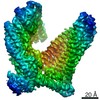
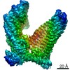
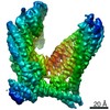
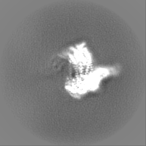
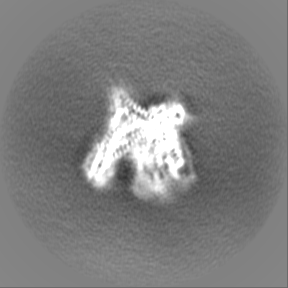
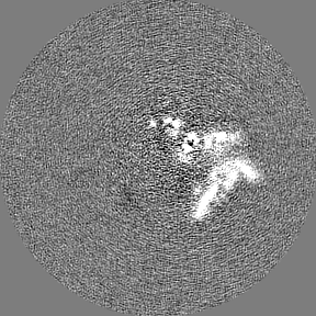
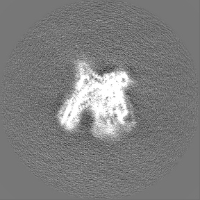
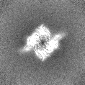
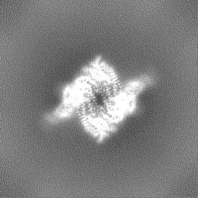
| 注釈 | Dimer, C2 reconstruction, unfiltered | ||||||||||||||||||||||||||||||||||||||||||||||||||||||||||||
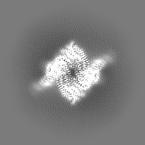
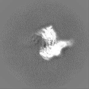
| 投影像・断面図 | 画像のコントロール
画像は Spider により作成 | ||||||||||||||||||||||||||||||||||||||||||||||||||||||||||||
| ボクセルのサイズ | X=Y=Z: 1.077 Å | ||||||||||||||||||||||||||||||||||||||||||||||||||||||||||||
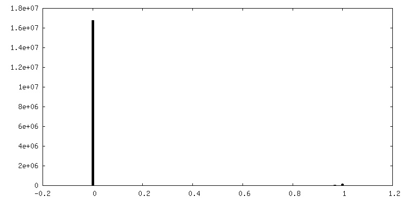
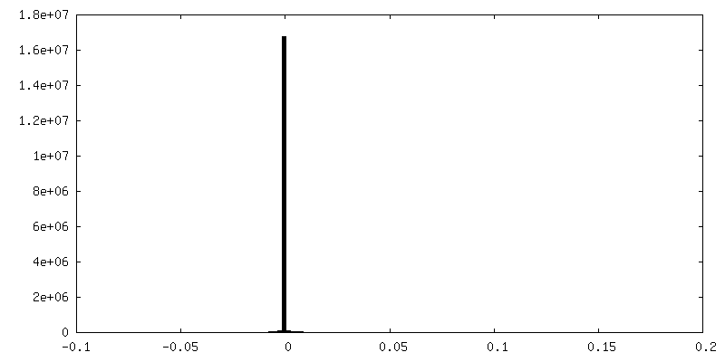
| 密度 |
| ||||||||||||||||||||||||||||||||||||||||||||||||||||||||||||
| 対称性 | 空間群: 1 | ||||||||||||||||||||||||||||||||||||||||||||||||||||||||||||
| 詳細 | EMDB XML:
CCP4マップ ヘッダ情報:
| ||||||||||||||||||||||||||||||||||||||||||||||||||||||||||||
 Z (Sec.)
 Y (Row.)
 X (Col.)


-添付データ
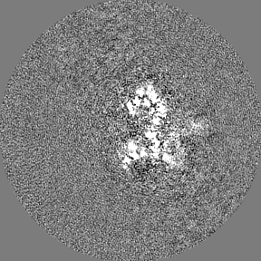
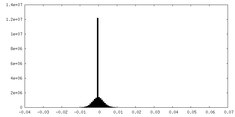
-マスク #1
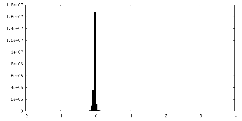
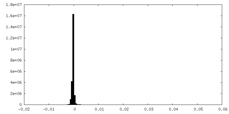
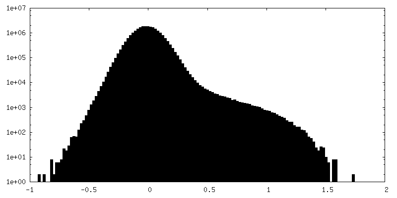
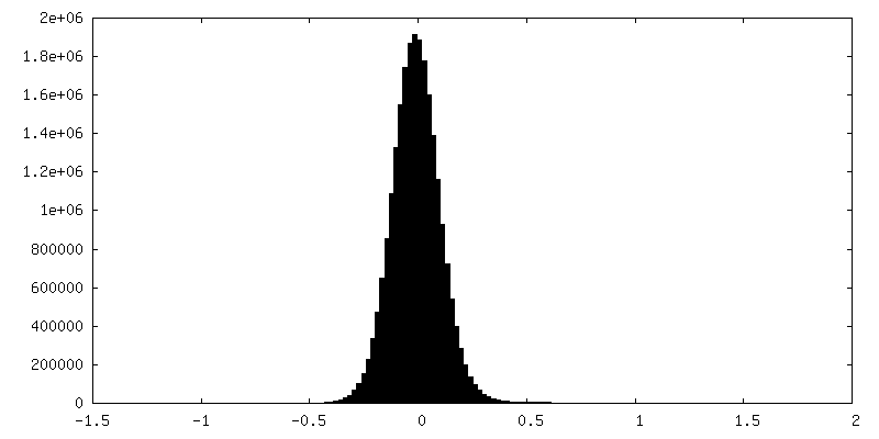
| ファイル | emd_11156_msk_1.map | ||||||||||||
|---|---|---|---|---|---|---|---|---|---|---|---|---|---|
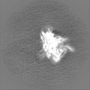
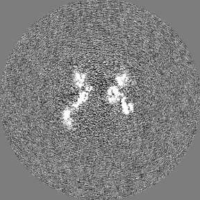
| 投影像・断面図 |
| ||||||||||||
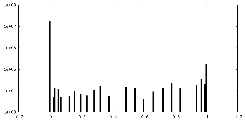
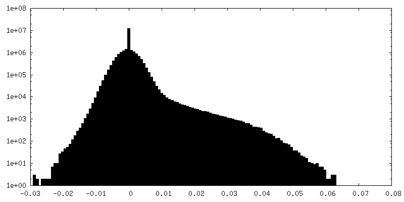
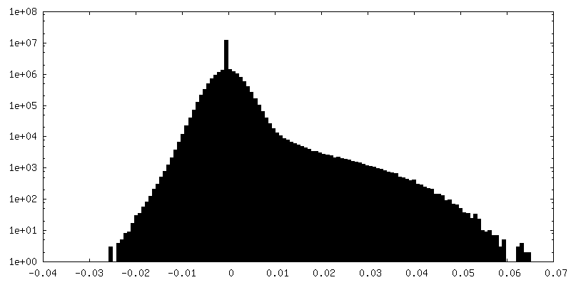
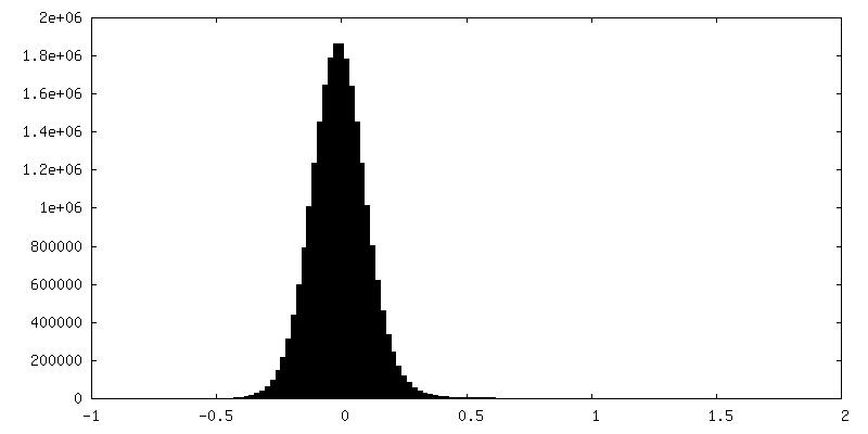
| 密度ヒストグラム |


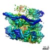
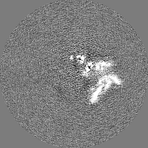
-追加マップ: Dimer, C2 reconstruction, sharpened
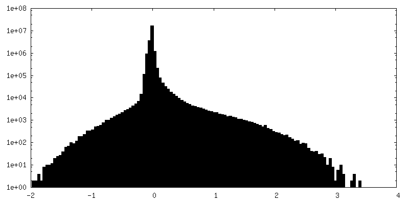
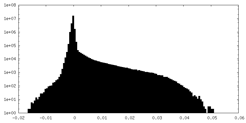
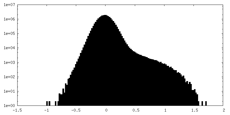
| ファイル | emd_11156_additional_1.map | ||||||||||||
|---|---|---|---|---|---|---|---|---|---|---|---|---|---|
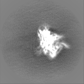
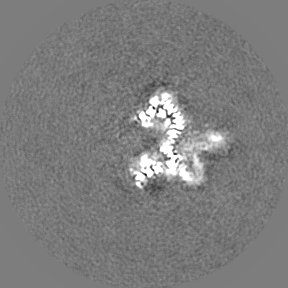
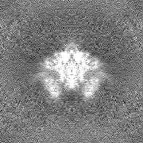
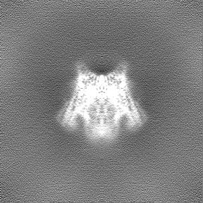
| 注釈 | Dimer, C2 reconstruction, sharpened | ||||||||||||
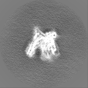
| 投影像・断面図 |
| ||||||||||||
| 密度ヒストグラム |

-追加マップ: C2 symmetry expansion, C1 monomer reconstruction, unfiltered
| ファイル | emd_11156_additional_2.map | ||||||||||||
|---|---|---|---|---|---|---|---|---|---|---|---|---|---|
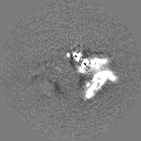
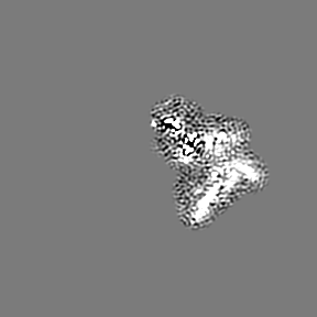
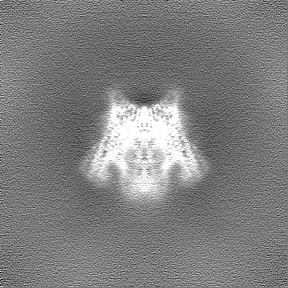
| 注釈 | C2 symmetry expansion, C1 monomer reconstruction, unfiltered | ||||||||||||
| 投影像・断面図 |
| ||||||||||||
| 密度ヒストグラム |
-追加マップ: C2 symmetry expansion, C1 monomer reconstruction, globally sharpened
| ファイル | emd_11156_additional_3.map | ||||||||||||
|---|---|---|---|---|---|---|---|---|---|---|---|---|---|
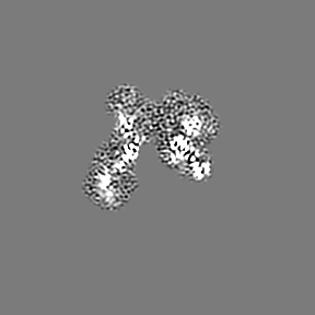
| 注釈 | C2 symmetry expansion, C1 monomer reconstruction, globally sharpened | ||||||||||||
| 投影像・断面図 |
| ||||||||||||
| 密度ヒストグラム |

-追加マップ: C2 symmetry expansion, C1 monomer reconstruction, half map 2
| ファイル | emd_11156_additional_4.map | ||||||||||||
|---|---|---|---|---|---|---|---|---|---|---|---|---|---|
| 注釈 | C2 symmetry expansion, C1 monomer reconstruction, half map 2 | ||||||||||||
| 投影像・断面図 |
| ||||||||||||
| 密度ヒストグラム |


-追加マップ: C2 symmetry expansion, C1 monomer reconstruction, half map 1
| ファイル | emd_11156_additional_5.map | ||||||||||||
|---|---|---|---|---|---|---|---|---|---|---|---|---|---|
| 注釈 | C2 symmetry expansion, C1 monomer reconstruction, half map 1 | ||||||||||||
| 投影像・断面図 |
| ||||||||||||
| 密度ヒストグラム |


-ハーフマップ: C2 dimer, half map 1
| ファイル | emd_11156_half_map_1.map | ||||||||||||
|---|---|---|---|---|---|---|---|---|---|---|---|---|---|
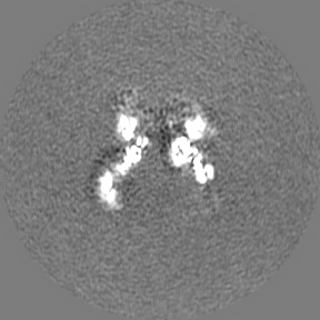
| 注釈 | C2 dimer, half map 1 | ||||||||||||
| 投影像・断面図 |
| ||||||||||||
| 密度ヒストグラム |
-ハーフマップ: C2 dimer, half map 2
| ファイル | emd_11156_half_map_2.map | ||||||||||||
|---|---|---|---|---|---|---|---|---|---|---|---|---|---|
| 注釈 | C2 dimer, half map 2 | ||||||||||||
| 投影像・断面図 |
| ||||||||||||
| 密度ヒストグラム |


- 試料の構成要素
-全体 : Merozoite surface protein 1 (MSP-1)
| 全体 | 名称: Merozoite surface protein 1 (MSP-1) |
|---|---|
| 要素 |
|
-超分子 #1: Merozoite surface protein 1 (MSP-1)
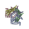
| 超分子 | 名称: Merozoite surface protein 1 (MSP-1) / タイプ: complex / ID: 1 / 親要素: 0 / 含まれる分子: all 詳細: Heteromeric assembly of p83/30 fusion and p38/42 fusion of MSP-1 from Plasmodium falciparum (dimer). |
|---|---|
| 由来(天然) | 生物種: |
-分子 #1: Precursor of the major merozoite surface antigens
| 分子 | 名称: Precursor of the major merozoite surface antigens / タイプ: protein_or_peptide / ID: 1 詳細: Fusion of the p83 and p30 subunits of the Merozoite surface protein-1 complex コピー数: 2 / 光学異性体: LEVO |
|---|---|
| 由来(天然) | 生物種: |
| 分子量 | 理論値: 101.973172 KDa |
| 組換発現 | 生物種:  |
| 配列 | 文字列: VTHESYQELV KKLEALEDAV LTGYSLFQKE KMVLNEEEIT TKGASAQSGA SAQSGASAQS GASAQSGASA QSGASAQSGT SGPSGPSGT SPSSRSNTLP RSNTSSGASP PADASDSDAK SYADLKHRVR NYLFTIKELK YPELFDLTNH MLTLCDNIHG F KYLIDGYE ...文字列: VTHESYQELV KKLEALEDAV LTGYSLFQKE KMVLNEEEIT TKGASAQSGA SAQSGASAQS GASAQSGASA QSGASAQSGT SGPSGPSGT SPSSRSNTLP RSNTSSGASP PADASDSDAK SYADLKHRVR NYLFTIKELK YPELFDLTNH MLTLCDNIHG F KYLIDGYE EINELLYKLN FYFDLLRAKL NDVCANDYCQ IPFNLKIRAN ELDVLKKLVF GYRKPLDNIK DNVGKMEDYI KK NKTTIAN INELIEGSKK TIDQNKNADN EEGKKKLYQA QYDLSIYNKQ LEEAHNLISV LEKRIDTLKK NENIKKLLDK INE IKNPPP ANSGNTPNTL LDKNKKIEEH EEKIKEIAKT IKFNIDSLFT DPLELEYYLR EKNKKVDVTP KSQDPTKSVQ IPKV PYPNG IVYPLPLTDI HNSLAADNDK NSYGDLMNPH TKEKINEKII TDNKERKIFI NNIKKKIDLE EKNINHTKEQ NKKLL EDYE KSKKDYEELL EKFYEMKFNN NFDKDVVDKI FSARYTYNVE KQRYNNKFSS SNNSVYNVQK LKKALSYLED YSLRKG ISE KDFNHYYTLK TGLEADIKKL TEEIKSSENK ILEKNFKGLT HSANGSLEVS DIVKLQVQKV LLIKKIEDLR KIELFLK NA QLKDSIHVPN IYKPQNKPEP YYLIVLKKEV DKLKEFIPKV KDMLKKEQAV LSSITQPLVA ASETTEDGGH STHTLSQS G ETEVTEETEE TEETVGHTTT VTITLPPTQP SPPKEVKVVE NSIEQKSNDN SQALTKTVYL KKLDEFLTKS YICHKYILV SNSSMDQKLL EVYNLTPEEE NELKSCDPLD LLFNIQNNIP AMYSLYDSMN NDLQHLFFEL YQKEMIYYLH KLKEENHIKK LLEEQKQIT GT UniProtKB: Merozoite surface protein 1 |
-分子 #2: Merozoite surface protein-1
| 分子 | 名称: Merozoite surface protein-1 / タイプ: protein_or_peptide / ID: 2 詳細: Fusion of the p38 and p42 subunits of the Merozoite surface protein-1 complex コピー数: 2 / 光学異性体: LEVO |
|---|---|
| 由来(天然) | 生物種: |
| 分子量 | 理論値: 89.615805 KDa |
| 組換発現 | 生物種: |
| 配列 | 文字列: SSTSSPGNTT VNTAQSATHS NSQNQQSNAS STNTQNGVAV SSGPAVVEES HDPLTVLSIS NDLKGIVSLL NLGNKTKVPN PLTISTTEM EKFYENILKN NDTYFNDDIK QFVKSNSKVI TGLTETQKNA LNDEIKKLKD TLQLSFDLYN KYKLKLDRLF N KKKELGQD ...文字列: SSTSSPGNTT VNTAQSATHS NSQNQQSNAS STNTQNGVAV SSGPAVVEES HDPLTVLSIS NDLKGIVSLL NLGNKTKVPN PLTISTTEM EKFYENILKN NDTYFNDDIK QFVKSNSKVI TGLTETQKNA LNDEIKKLKD TLQLSFDLYN KYKLKLDRLF N KKKELGQD KMQIKKLTLL KEQLESKLNS LNNPHNVLQN FSVFFNKKKE AEIAETENTL ENTKILLKHY KGLVKYYNGE SS PLKTLSE VSIQTEDNYA NLEKFRVLSK IDGKLNDNLH LGKKKLSFLS SGLHHLITEL KEVIKNKNYT GNSPSENNKK VNE ALKSYE NFLPEAKVTT VVTPPQPDVT PSPLSVRVSG SSGSTKEETQ IPTSGSLLTE LQQVVQLQNY DEEDDSLVVL PIFG ESEDN DEYLDQVVTG EAISVTMDNI LSGFENEYDV IYLKPLAGVY RSLKKQIEKN IFTFNLNLND ILNSRLKKRK YFLDV LESD LMQFKHISSN EYIIEDSFKL LNSEQKNTLL KSYKYIKESV ENDIKFAQEG ISYYEKVLAK YKDDLESIKK VIKEEK EKF PSSPPTTPPS PAKTDEQKKE SKFLPFLTNI ETLYNNLVNK IDDYLINLKA KINDCNVEKD EAHVKITKLS DLKAIDD KI DLFKNPYDFE AIKKLINDDT KKDMLGKLLS TGLVQNFPNT IISKLIEGKF QDMLNISQHQ CVKKQCPENS GCFRHLDE R EECKCLLNYK QEGDKCVENP NPTCNENNGG CDADATCTEE DSGSSRKKIT CECTKPDSYP LFDGIFCSSS N UniProtKB: Merozoite surface protein 1 |
-実験情報
-構造解析
| 手法 | クライオ電子顕微鏡法 |
|---|---|
 解析 解析 | 単粒子再構成法 |
| 試料の集合状態 | particle |
-試料調製
| 緩衝液 | pH: 7.4 |
|---|---|
| 凍結 | 凍結剤: ETHANE |
- 電子顕微鏡法
| 顕微鏡 | FEI TITAN KRIOS |
|---|---|
| 撮影 | フィルム・検出器のモデル: GATAN K2 SUMMIT (4k x 4k) 平均電子線量: 60.0 e/Å2 |
| 電子線 | 加速電圧: 300 kV / 電子線源:  FIELD EMISSION GUN FIELD EMISSION GUN |
| 電子光学系 | 照射モード: FLOOD BEAM / 撮影モード: BRIGHT FIELD / 最大 デフォーカス(公称値): 3.0 µm / 最小 デフォーカス(公称値): 0.5 µm / 倍率(公称値): 130000 |
| 試料ステージ | 試料ホルダーモデル: FEI TITAN KRIOS AUTOGRID HOLDER ホルダー冷却材: NITROGEN |
| 実験機器 |  モデル: Titan Krios / 画像提供: FEI Company |