 Movie
Movie Controller

[English] 日本語
 Yorodumi
- EMDB-1965: Structural and Functional Studies of LRP6 Ectodomain Reveal a Pla... -
+ Open data

- Basic information
| Entry | Database: EMDB / ID: EMD-1965 | |||||||||
|---|---|---|---|---|---|---|---|---|---|---|
| Title | Structural and Functional Studies of LRP6 Ectodomain Reveal a Platform for Wnt Signaling | |||||||||
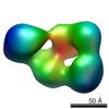
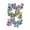
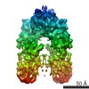
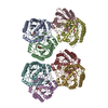
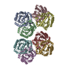
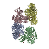
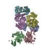
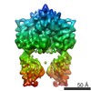
 Map data Map data | This is a surface rendering of the ectodomain of LRP6 bound to its chaperone Mesd | |||||||||
 Sample Sample |
| |||||||||
 Keywords Keywords | LDL-receptor-related protein 6 / Wnt signaling pathway / Wnt co-receptor | |||||||||
| Biological species |  Homo sapiens (human) Homo sapiens (human) | |||||||||
| Method | single particle reconstruction / negative staining / Resolution: 26.0 Å | |||||||||
 Authors Authors | Chen S / Bubeck D / MacDonald BT / Liang WX / Mao JH / Malinauskas T / Llorca O / Aricescu AR / Siebold C / He X / Jones EY | |||||||||
 Citation Citation | Journal: Dev Cell / Year: 2011 Title: Structural and functional studies of LRP6 ectodomain reveal a platform for Wnt signaling. Authors: Shuo Chen / Doryen Bubeck / Bryan T MacDonald / Wen-Xue Liang / Jian-Hua Mao / Tomas Malinauskas / Oscar Llorca / A Radu Aricescu / Christian Siebold / Xi He / E Yvonne Jones /  Abstract: LDL-receptor-related protein 6 (LRP6), alongside Frizzled receptors, transduces Wnt signaling across the plasma membrane. The LRP6 ectodomain comprises four tandem β-propeller-EGF-like domain (PE) ...LDL-receptor-related protein 6 (LRP6), alongside Frizzled receptors, transduces Wnt signaling across the plasma membrane. The LRP6 ectodomain comprises four tandem β-propeller-EGF-like domain (PE) pairs that harbor binding sites for Wnt morphogens and their antagonists including Dickkopf 1 (Dkk1). To understand how these multiple interactions are integrated, we combined crystallographic analysis of the third and fourth PE pairs with electron microscopy (EM) to determine the complete ectodomain structure. An extensive inter-pair interface, conserved for the first-to-second and third-to-fourth PE interactions, contributes to a compact platform-like architecture, which is disrupted by mutations implicated in developmental diseases. EM reconstruction of the LRP6 platform bound to chaperone Mesd exemplifies a binding mode spanning PE pairs. Cellular and binding assays identify overlapping Wnt3a- and Dkk1-binding surfaces on the third PE pair, consistent with steric competition, but also suggest a model in which the platform structure supports an interplay of ligands through multiple interaction sites. | |||||||||
| History |
|
- Structure visualization
| Movie |
 Movie viewer Movie viewer |
|---|---|
| Structure viewer | EM map: SurfViewMolmilJmol/JSmol |
| Supplemental images |
 UCSF Chimera
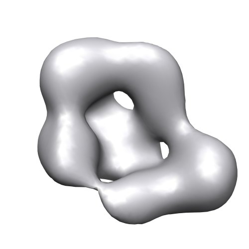
- Downloads & links
-EMDB archive
| Map data | emd_1965.map.gz | 790.7 KB | EMDB map data format | |
|---|---|---|---|---|
| Header (meta data) | emd-1965-v30.xmlemd-1965.xml | 9.7 KB 9.7 KB | Display Display | EMDB header |
| Images |  1965_LRP6_MESD3.jpg 1965_LRP6_MESD3.jpg | 42.4 KB | ||
| Archive directory |  http://ftp.pdbj.org/pub/emdb/structures/EMD-1965ftp://ftp.pdbj.org/pub/emdb/structures/EMD-1965 http://ftp.pdbj.org/pub/emdb/structures/EMD-1965ftp://ftp.pdbj.org/pub/emdb/structures/EMD-1965 | HTTPS FTP |
-Validation report
| Summary document | emd_1965_validation.pdf.gz | 197.5 KB | Display | EMDB validaton report |
|---|---|---|---|---|
| Full document | emd_1965_full_validation.pdf.gz | 196.6 KB | Display | |
| Data in XML | emd_1965_validation.xml.gz | 4.7 KB | Display | |
| Arichive directory | https://ftp.pdbj.org/pub/emdb/validation_reports/EMD-1965ftp://ftp.pdbj.org/pub/emdb/validation_reports/EMD-1965 | HTTPS FTP |
-Related structure data
-Links
| EMDB pages | EMDB (EBI/PDBe) / EMDataResource |
|---|
-Map
| File | Download / File: emd_1965.map.gz / Format: CCP4 / Size: 825.2 KB / Type: IMAGE STORED AS FLOATING POINT NUMBER (4 BYTES) | ||||||||||||||||||||||||||||||||||||||||||||||||||||||||||||||||||||
|---|---|---|---|---|---|---|---|---|---|---|---|---|---|---|---|---|---|---|---|---|---|---|---|---|---|---|---|---|---|---|---|---|---|---|---|---|---|---|---|---|---|---|---|---|---|---|---|---|---|---|---|---|---|---|---|---|---|---|---|---|---|---|---|---|---|---|---|---|---|
| Annotation | This is a surface rendering of the ectodomain of LRP6 bound to its chaperone Mesd | ||||||||||||||||||||||||||||||||||||||||||||||||||||||||||||||||||||
| Voxel size | X=Y=Z: 4.56 Å | ||||||||||||||||||||||||||||||||||||||||||||||||||||||||||||||||||||
| Density |
| ||||||||||||||||||||||||||||||||||||||||||||||||||||||||||||||||||||
| Symmetry | Space group: 1 | ||||||||||||||||||||||||||||||||||||||||||||||||||||||||||||||||||||
| Details | EMDB XML:
CCP4 map header:
| ||||||||||||||||||||||||||||||||||||||||||||||||||||||||||||||||||||
-Supplemental data
- Sample components
-Entire : Ectodomain of LRP6 residues 20 to 1361 and chaperone Mesd
| Entire | Name: Ectodomain of LRP6 residues 20 to 1361 and chaperone Mesd |
|---|---|
| Components |
|
-Supramolecule #1000: Ectodomain of LRP6 residues 20 to 1361 and chaperone Mesd
| Supramolecule | Name: Ectodomain of LRP6 residues 20 to 1361 and chaperone Mesd type: sample / ID: 1000 / Details: The sample was monodisperse / Oligomeric state: one LRP6 binds to one Mesd / Number unique components: 2 |
|---|---|
| Molecular weight | Experimental: 200 KDa / Method: Multi angle light scattering |
-Macromolecule #1: LDL-receptor-related protein 6
| Macromolecule | Name: LDL-receptor-related protein 6 / type: protein_or_peptide / ID: 1 / Name.synonym: LRP6 / Number of copies: 1 / Oligomeric state: Monomer / Recombinant expression: Yes |
|---|---|
| Source (natural) | Organism: Homo sapiens (human) / synonym: Human |
| Molecular weight | Experimental: 100 KDa |
| Recombinant expression | Organism: HEK293S / Recombinant plasmid: pHLsec vector |
-Macromolecule #2: Mesoderm development protein
| Macromolecule | Name: Mesoderm development protein / type: protein_or_peptide / ID: 2 / Name.synonym: Mesd / Number of copies: 1 / Oligomeric state: Monomer / Recombinant expression: Yes |
|---|---|
| Source (natural) | Organism: Homo sapiens (human) / synonym: Human |
| Molecular weight | Experimental: 100 KDa |
| Recombinant expression | Organism: HEK293S / Recombinant plasmid: pHLsec vector |
-Experimental details
-Structure determination
| Method | negative staining |
|---|---|
 Processing Processing | single particle reconstruction |
| Aggregation state | particle |
-Sample preparation
| Concentration | 0.015 mg/mL |
|---|---|
| Buffer | pH: 8 / Details: 150 mM NaCl,10 mM Tris-HCl |
| Staining | Type: NEGATIVE Details: Grids were negatively stained with 0.75% uranyl formate using the two-drop method |
| Grid | Details: carbon-coated copper grids |
| Vitrification | Cryogen name: NONE / Instrument: OTHER |
- Electron microscopy
| Microscope | JEOL 1230 |
|---|---|
| Image recording | Category: CCD / Film or detector model: TVIPS TEMCAM-F416 (4k x 4k) / Digitization - Sampling interval: 4.56 µm / Average electron dose: 10 e/Å2 |
| Electron beam | Acceleration voltage: 100 kV / Electron source: LAB6 |
| Electron optics | Illumination mode: FLOOD BEAM / Imaging mode: BRIGHT FIELD / Cs: 2.9 mm / Nominal magnification: 72500 |
| Sample stage | Specimen holder: single-tilt / Specimen holder model: JEOL |
-Image processing
| Final reconstruction | Applied symmetry - Point group: C1 (asymmetric) / Algorithm: OTHER / Resolution.type: BY AUTHOR / Resolution: 26.0 Å / Resolution method: FSC 0.5 CUT-OFF / Software - Name: EMAN, XMIPP / Number images used: 7928 |
|---|
