 Movie
Movie Controller

[English] 日本語
 Yorodumi
- PDB-1hfb: Crystal structure of the tyrosine-regulated 3-deoxy-D-arabino-hep... -
+ Open data

- Basic information
| Entry | Database: PDB / ID: 1hfb | ||||||
|---|---|---|---|---|---|---|---|
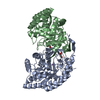
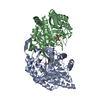
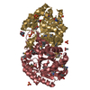
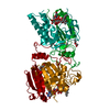
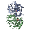
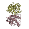
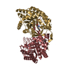
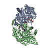
| Title | Crystal structure of the tyrosine-regulated 3-deoxy-D-arabino-heptulosonate-7-phosphate synthase from Saccharomyces cerevisiae complexed with phosphoenolpyruvate | ||||||
 Components Components | TYROSINE-REGULATED 3-DEOXY-D-ARABINO-HEPTULOSONATE-7-PHOSPHATE SYNTHASE | ||||||
 Keywords Keywords |  LYASE / BETA-ALPHA-BARREL LYASE / BETA-ALPHA-BARREL | ||||||
| Function / homology |  Function and homology information3-deoxy-7-phosphoheptulonate synthase / 3-deoxy-7-phosphoheptulonate synthase activity / chorismate biosynthetic process / aromatic amino acid family biosynthetic process / amino acid biosynthetic process / nucleus / cytoplasm Function and homology information3-deoxy-7-phosphoheptulonate synthase / 3-deoxy-7-phosphoheptulonate synthase activity / chorismate biosynthetic process / aromatic amino acid family biosynthetic process / amino acid biosynthetic process / nucleus / cytoplasmSimilarity search - Function | ||||||
| Biological species |  SACCHAROMYCES CEREVISIAE (brewer's yeast) SACCHAROMYCES CEREVISIAE (brewer's yeast) | ||||||
| Method | X-RAY DIFFRACTION / SYNCHROTRON / MOLECULAR REPLACEMENT / Resolution: 1.9 Å | ||||||
 Authors Authors | Schneider, T.R. / Hartmann, M. / Braus, G.H. | ||||||
 Citation Citation | Journal: Proc.Natl.Acad.Sci.USA / Year: 2003 Title: Evolution of Feedback-Inhibited Beta /Alpha Barrel Isoenzymes by Gene Duplication and a Single Mutation Authors: Hartmann, M. / Schneider, T.R. / Pfeil, A. / Heinrich, G. / Lipscomb, W. / Braus, G.H. | ||||||
| History |
| ||||||
| Remark 700 | SHEET DETERMINATION METHOD: DSSP THE SHEETS PRESENTED AS AB, BA, CB, DA, EB, FA, GB, AND HA BELOW ... SHEET DETERMINATION METHOD: DSSP THE SHEETS PRESENTED AS AB, BA, CB, DA, EB, FA, GB, AND HA BELOW ARE ACTUALLY 8-STRANDED BARRELS. THESE ARE REPRESENTED BY A 9-STRANDED SHEET IN WHICH THE FIRST AND LAST STRANDS ARE IDENTICAL. |
- Structure visualization
| Structure viewer | Molecule: MolmilJmol/JSmol |
|---|
- Downloads & links
-Download
| PDBx/mmCIF format | 1hfb.cif.gz | 530 KB | Display | PDBx/mmCIF format |
|---|---|---|---|---|
| PDB format | pdb1hfb.ent.gz | 436.8 KB | Display | PDB format |
| PDBx/mmJSON format | 1hfb.json.gz | Tree view | PDBx/mmJSON format | |
| Others |  Other downloads Other downloads |
-Validation report
| Arichive directory | https://data.pdbj.org/pub/pdb/validation_reports/hf/1hfbftp://data.pdbj.org/pub/pdb/validation_reports/hf/1hfb | HTTPS FTP |
|---|
-Related structure data
| Related structure data | 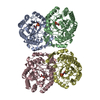 1qr7S S: Starting model for refinement |
|---|---|
| Similar structure data |


-Links
 PDBj
PDBj
- Assembly
| Deposited unit | 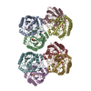
| ||||||||
|---|---|---|---|---|---|---|---|---|---|
| 1 | 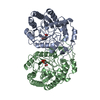
| ||||||||
| 2 | 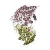
| ||||||||
| 3 | 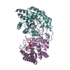
| ||||||||
| 4 | 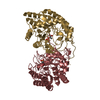
| ||||||||
| Unit cell |
| ||||||||
| Noncrystallographic symmetry (NCS) | NCS oper: (Code: given / Matrix: (1),| Details | THE 8 MOLECULES CONTAINED IN THE ASSYMETRIC UNIT AREARRANGED IN TWO TETRAMERS (DIMER OF DIMERS) WITH APPROXIMATE222 POINT SYMMETRY. THE TETRAMERS HAVE VERY SIMILAR ORIENTATIONS,WITH THEIR CENTERS OF MASS BEING RELATED BY ACRYSTALLOGRAPHIC TRANSLATION OF (0.5,0.5,0). | |
-Components
| #1: Protein | Mass: 39797.055 Da / Num. of mol.: 8 Source method: isolated from a genetically manipulated source Source: (gene. exp.) SACCHAROMYCES CEREVISIAE (brewer's yeast)Strain: RH1326 / Production host: SACCHAROMYCES CEREVISIAE (brewer's yeast) / Strain (production host): RH1326 / References: UniProt: P32449, EC: 4.1.2.15#2: Chemical | ChemComp-PEP / Phosphoenolpyruvic acid  Mass: 168.042 Da / Num. of mol.: 8 / Source method: obtained synthetically / Formula: C3H5O6P Mass: 168.042 Da / Num. of mol.: 8 / Source method: obtained synthetically / Formula: C3H5O6P#3: Water | ChemComp-HOH / | Water Mass: 18.015 Da / Num. of mol.: 1251 / Source method: isolated from a natural source / Formula: H2O Mass: 18.015 Da / Num. of mol.: 1251 / Source method: isolated from a natural source / Formula: H2O |
|---|
-Experimental details
-Experiment
| Experiment | Method: X-RAY DIFFRACTION / Number of used crystals: 1 |
|---|
- Sample preparation
| Crystal | Density Matthews: 2.22 Å3/Da / Density % sol: 44.69 % | ||||||||||||||||||||||||
|---|---|---|---|---|---|---|---|---|---|---|---|---|---|---|---|---|---|---|---|---|---|---|---|---|---|
| Crystal grow | pH: 9 / Details: pH 9.00 | ||||||||||||||||||||||||
| Crystal grow | *PLUS Temperature: 21 ℃ / Method: vapor diffusion, hanging drop | ||||||||||||||||||||||||
| Components of the solutions | *PLUS
|
-Data collection
| Diffraction | Mean temperature: 100 K |
|---|---|
| Diffraction source | Source: SYNCHROTRON / Site: EMBL/DESY, HAMBURG  / Beamline: X11 / Wavelength: 0.9076 / Beamline: X11 / Wavelength: 0.9076 |
| Detector | Type: MARRESEARCH / Detector: IMAGE PLATE / Date: Sep 1, 1998 |
| Radiation | Protocol: SINGLE WAVELENGTH / Monochromatic (M) / Laue (L): M / Scattering type: x-ray |
| Radiation wavelength | Wavelength: 0.9076 Å / Relative weight: 1 |
| Reflection | Resolution: 1.9→40 Å / Num. obs: 210365 / % possible obs: 96.3 % / Observed criterion σ(I): -3 / Redundancy: 2.01 % / Rmerge(I) obs: 0.037 |
| Reflection | *PLUS Redundancy: 2.9 % / Num. measured all: 423490 |
| Reflection shell | *PLUS Highest resolution: 1.9 Å / Lowest resolution: 1.97 Å / % possible obs: 89.3 % / Redundancy: 1.2 % / Rmerge(I) obs: 0.354 / Mean I/σ(I) obs: 2.2 |
- Processing
| Software |
| ||||||||||||||||||||||||||||||||||||||||||||||||||||||||||||||||||||||||||||||||
|---|---|---|---|---|---|---|---|---|---|---|---|---|---|---|---|---|---|---|---|---|---|---|---|---|---|---|---|---|---|---|---|---|---|---|---|---|---|---|---|---|---|---|---|---|---|---|---|---|---|---|---|---|---|---|---|---|---|---|---|---|---|---|---|---|---|---|---|---|---|---|---|---|---|---|---|---|---|---|---|---|---|
| Refinement | Method to determine structure: MOLECULAR REPLACEMENT Starting model: CHAINS A AND B OF PDB ENTRY 1QR7 Resolution: 1.9→40 Å / Data cutoff high absF: 10000 / Cross valid method: THROUGHOUT / σ(F): 0
| ||||||||||||||||||||||||||||||||||||||||||||||||||||||||||||||||||||||||||||||||
| Solvent computation | Solvent model: FLAT MODEL / Bsol: 41.9802 Å2 / ksol: 0.34361 e/Å3 | ||||||||||||||||||||||||||||||||||||||||||||||||||||||||||||||||||||||||||||||||
| Displacement parameters |
| ||||||||||||||||||||||||||||||||||||||||||||||||||||||||||||||||||||||||||||||||
| Refinement step | Cycle: LAST / Resolution: 1.9→40 Å
| ||||||||||||||||||||||||||||||||||||||||||||||||||||||||||||||||||||||||||||||||
| Refine LS restraints |
| ||||||||||||||||||||||||||||||||||||||||||||||||||||||||||||||||||||||||||||||||
| Xplor file |
| ||||||||||||||||||||||||||||||||||||||||||||||||||||||||||||||||||||||||||||||||
| Refinement | *PLUS Rfactor Rfree: 0.261 / Rfactor Rwork: 0.208 | ||||||||||||||||||||||||||||||||||||||||||||||||||||||||||||||||||||||||||||||||
| Solvent computation | *PLUS | ||||||||||||||||||||||||||||||||||||||||||||||||||||||||||||||||||||||||||||||||
| Displacement parameters | *PLUS | ||||||||||||||||||||||||||||||||||||||||||||||||||||||||||||||||||||||||||||||||
| Refine LS restraints | *PLUS
|
