 Movie
Movie Controller

[English] 日本語
 Yorodumi
- PDB-3jax: Heavy meromyosin from Schistosoma mansoni muscle thick filament b... -
+ Open data

- Basic information
| Entry | Database: PDB / ID: 3jax | ||||||
|---|---|---|---|---|---|---|---|
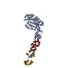
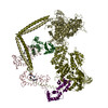
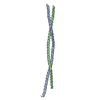
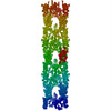
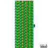
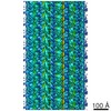
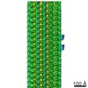
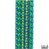
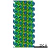
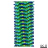
| Title | Heavy meromyosin from Schistosoma mansoni muscle thick filament by negative stain EM | ||||||
 Components Components |
| ||||||
 Keywords Keywords | CONTRACTILE PROTEIN / muscle protein / smooth muscle / myosin subfragment 2 / heavy meromyosin / essential light chain / regulatory light chain / motor protein / coiled-coil | ||||||
| Function / homology |  Function and homology information Function and homology informationmyosin complex / cytoskeletal motor activity / actin filament binding / calcium ion binding / ATP binding Similarity search - Function | ||||||
| Biological species |  | ||||||
| Method | ELECTRON MICROSCOPY / helical reconstruction / negative staining / Resolution: 23 Å | ||||||
 Authors Authors | Sulbaran, G. / Alamo, L. / Pinto, A. / Marquez, G. / Mendez, F. / Padron, R. / Craig, R. | ||||||
 Citation Citation | Journal: Proc Natl Acad Sci U S A / Year: 2015 Title: An invertebrate smooth muscle with striated muscle myosin filaments. Authors: Guidenn Sulbarán / Lorenzo Alamo / Antonio Pinto / Gustavo Márquez / Franklin Méndez / Raúl Padrón / Roger Craig /   Abstract: Muscle tissues are classically divided into two major types, depending on the presence or absence of striations. In striated muscles, the actin filaments are anchored at Z-lines and the myosin and ...Muscle tissues are classically divided into two major types, depending on the presence or absence of striations. In striated muscles, the actin filaments are anchored at Z-lines and the myosin and actin filaments are in register, whereas in smooth muscles, the actin filaments are attached to dense bodies and the myosin and actin filaments are out of register. The structure of the filaments in smooth muscles is also different from that in striated muscles. Here we have studied the structure of myosin filaments from the smooth muscles of the human parasite Schistosoma mansoni. We find, surprisingly, that they are indistinguishable from those in an arthropod striated muscle. This structural similarity is supported by sequence comparison between the schistosome myosin II heavy chain and known striated muscle myosins. In contrast, the actin filaments of schistosomes are similar to those of smooth muscles, lacking troponin-dependent regulation. We conclude that schistosome muscles are hybrids, containing striated muscle-like myosin filaments and smooth muscle-like actin filaments in a smooth muscle architecture. This surprising finding has broad significance for understanding how muscles are built and how they evolved, and challenges the paradigm that smooth and striated muscles always have distinctly different components. #1: Journal: J Mol Biol / Year: 2008Title: Three-dimensional reconstruction of tarantula myosin filaments suggests how phosphorylation may regulate myosin activity. Authors: Lorenzo Alamo / Willy Wriggers / Antonio Pinto / Fulvia Bártoli / Leiria Salazar / Fa-Qing Zhao / Roger Craig / Raúl Padrón / Abstract: Muscle contraction involves the interaction of the myosin heads of the thick filaments with actin subunits of the thin filaments. Relaxation occurs when this interaction is blocked by molecular ...Muscle contraction involves the interaction of the myosin heads of the thick filaments with actin subunits of the thin filaments. Relaxation occurs when this interaction is blocked by molecular switches on these filaments. In many muscles, myosin-linked regulation involves phosphorylation of the myosin regulatory light chains (RLCs). Electron microscopy of vertebrate smooth muscle myosin molecules (regulated by phosphorylation) has provided insight into the relaxed structure, revealing that myosin is switched off by intramolecular interactions between its two heads, the free head and the blocked head. Three-dimensional reconstruction of frozen-hydrated specimens revealed that this asymmetric head interaction is also present in native thick filaments of tarantula striated muscle. Our goal in this study was to elucidate the structural features of the tarantula filament involved in phosphorylation-based regulation. A new reconstruction revealed intra- and intermolecular myosin interactions in addition to those seen previously. To help interpret the interactions, we sequenced the tarantula RLC and fitted an atomic model of the myosin head that included the predicted RLC atomic structure and an S2 (subfragment 2) crystal structure to the reconstruction. The fitting suggests one intramolecular interaction, between the cardiomyopathy loop of the free head and its own S2, and two intermolecular interactions, between the cardiac loop of the free head and the essential light chain of the blocked head and between the Leu305-Gln327 interaction loop of the free head and the N-terminal fragment of the RLC of the blocked head. These interactions, added to those previously described, would help switch off the thick filament. Molecular dynamics simulations suggest how phosphorylation could increase the helical content of the RLC N-terminus, weakening these interactions, thus releasing both heads and activating the thick filament. #2: Journal: J Mol Biol / Year: 2003 Title: Refined model of the 10S conformation of smooth muscle myosin by cryo-electron microscopy 3D image reconstruction. Authors: Jun Liu / Thomas Wendt / Dianne Taylor / Kenneth Taylor / Abstract: The actin-activated ATPase activity of smooth muscle myosin and heavy meromyosin (smHMM) is regulated by phosphorylation of the regulatory light chain (RLC). Complete regulation requires two intact ...The actin-activated ATPase activity of smooth muscle myosin and heavy meromyosin (smHMM) is regulated by phosphorylation of the regulatory light chain (RLC). Complete regulation requires two intact myosin heads because single-headed myosin subfragments are always active. 2D crystalline arrays of the 10S form of intact myosin, which has a dephosphorylated RLC, were produced on a positively charged lipid monolayer and imaged in 3D at 2.0 nm resolution by cryo-electron microscopy of frozen, hydrated specimens. An atomic model of smooth muscle myosin was constructed from the X-ray structures of the smooth muscle myosin motor domain and essential light chain and a homology model of the RLC was produced based on the skeletal muscle S1 structure. The initial model of the 10S myosin, based on the previous reconstruction of smHMM, was subjected to real space refinement to obtain a quantitative fit to the density. The smHMM was likewise refined and both refined models reveal the same asymmetric interaction between the upper 50 kDa domain of the "blocked" head and parts of the catalytic, converter domains and the essential light chain of the "free" head observed previously. This observation suggests that this interaction is not simply due to crystallographic packing but is enforced by elements of the myosin heads. The 10S reconstruction shows additional alpha-helical coiled-coil not seen in the earlier smHMM reconstruction, but the location of one segment of S2 is the same in both. #3: Journal: Proc Natl Acad Sci U S A / Year: 2006Title: Crystal structures of human cardiac beta-myosin II S2-Delta provide insight into the functional role of the S2 subfragment. Authors: Wulf Blankenfeldt / Nicolas H Thomä / John S Wray / Mathias Gautel / Ilme Schlichting /  Abstract: Myosin II is the major component of the muscle thick filament. It consists of two N-terminal S1 subfragments ("heads") connected to a long dimeric coiled-coil rod. The rod is in itself twofold ...Myosin II is the major component of the muscle thick filament. It consists of two N-terminal S1 subfragments ("heads") connected to a long dimeric coiled-coil rod. The rod is in itself twofold symmetric, but in the filament, the two heads point away from the filament surface and are therefore not equivalent. This breaking of symmetry requires the initial section of the rod, subfragment 2 (S2), to be relatively flexible. S2 is an important functional element, involved in various mechanisms by which the activity of smooth and striated muscle is regulated. We have determined crystal structures of the 126 N-terminal residues of S2 from human cardiac beta-myosin II (S2-Delta), of both WT and the disease-associated E924K mutant. S2-Delta is a straight parallel dimeric coiled coil, but the N terminus of one chain is disordered in WT-S2-Delta due to crystal contacts, indicative of unstable local structure. Bulky noncanonical side chains pack into a/d positions of S2-Delta's N terminus, leading to defined local asymmetry and axial stagger, which could induce nonequivalence of the S1 subfragments. Additionally, S2 possesses a conserved charge distribution with three prominent rings of negative potential within S2-Delta, the first of which may provide a binding interface for the "blocked head" of smooth muscle myosin in the OFF state. The observation that many disease-associated mutations affect the second negatively charged ring further suggests that charge interactions play an important role in regulation of cardiac muscle activity through myosin-binding protein C. #4: Journal: Cell / Year: 1999Title: Atomic structure of scallop myosin subfragment S1 complexed with MgADP: a novel conformation of the myosin head. Authors: A Houdusse / V N Kalabokis / D Himmel / A G Szent-Györgyi / C Cohen / Abstract: The crystal structure of a proteolytic subfragment from scallop striated muscle myosin, complexed with MgADP, has been solved at 2.5 A resolution and reveals an unusual conformation of the myosin ...The crystal structure of a proteolytic subfragment from scallop striated muscle myosin, complexed with MgADP, has been solved at 2.5 A resolution and reveals an unusual conformation of the myosin head. The converter and the lever arm are in very different positions from those in either the pre-power stroke or near-rigor state structures; moreover, in contrast to these structures, the SH1 helix is seen to be unwound. Here we compare the overall organization of the myosin head in these three states and show how the conformation of three flexible "joints" produces rearrangements of the four major subdomains in the myosin head with different bound nucleotides. We believe that this novel structure represents one of the prehydrolysis ("ATP") states of the contractile cycle in which the myosin heads stay detached from actin. | ||||||
| History |
|
- Structure visualization
| Movie |
Movie viewer |
|---|---|
| Structure viewer | Molecule: MolmilJmol/JSmol |
 UCSF Chimera
- Downloads & links
-Download
| PDBx/mmCIF format | 3jax.cif.gz | 539 KB | Display | PDBx/mmCIF format |
|---|---|---|---|---|
| PDB format | pdb3jax.ent.gz | 445.8 KB | Display | PDB format |
| PDBx/mmJSON format | 3jax.json.gz | Tree view | PDBx/mmJSON format | |
| Others |  Other downloads Other downloads |
-Validation report
| Summary document | 3jax_validation.pdf.gz | 745.5 KB | Display | wwPDB validaton report |
|---|---|---|---|---|
| Full document | 3jax_full_validation.pdf.gz | 852.7 KB | Display | |
| Data in XML | 3jax_validation.xml.gz | 79.9 KB | Display | |
| Data in CIF | 3jax_validation.cif.gz | 120.2 KB | Display | |
| Arichive directory | https://data.pdbj.org/pub/pdb/validation_reports/ja/3jaxftp://data.pdbj.org/pub/pdb/validation_reports/ja/3jax | HTTPS FTP |
-Related structure data
| Related structure data | 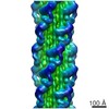 6370MC M: map data used to model this data C: citing same article ( |
|---|---|
| Similar structure data |
-Links
 PDBj
PDBj


- Assembly
| Deposited unit | 
|
|---|---|
| 1 | x 20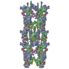
|
| 2 |
|
| 3 | 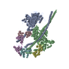
|
| Symmetry | Helical symmetry: (Circular symmetry: 4 / Dyad axis: no / N subunits divisor: 1 / Num. of operations: 20 / Rise per n subunits: 145 Å / Rotation per n subunits: 30 °) |
-Components
| #1: Protein | Mass: 112182.516 Da / Num. of mol.: 2 / Source method: isolated from a natural source / Source: (natural) #2: Protein | Mass: 17004.221 Da / Num. of mol.: 2 / Source method: isolated from a natural source / Source: (natural) #3: Protein | Mass: 21709.250 Da / Num. of mol.: 2 / Source method: isolated from a natural source / Source: (natural) Sequence details | THE IMAGED FILAMENTS ARE FROM SCHISTOSOMA MANSONI, BUT THE MODELED SEQUENCES ARE FROM CHICKEN/HUMAN ...THE IMAGED FILAMENTS ARE FROM SCHISTOSOM | |
|---|
-Experimental details
-Experiment
| Experiment | Method: ELECTRON MICROSCOPY |
|---|---|
| EM experiment | Aggregation state: FILAMENT / 3D reconstruction method: helical reconstruction |
- Sample preparation
| Component |
| |||||||||||||||
|---|---|---|---|---|---|---|---|---|---|---|---|---|---|---|---|---|
| Buffer solution | Name: 100 mM NaCl, 3 mM MgCl2, 1 mM EGTA, 5 mM PIPES, 1mM NaN3, 5 mM MgATP, 0.01 mM blebbistatin, protease inhibitor cocktail (Sigma P-8465) pH: 7 Details: 100 mM NaCl, 3 mM MgCl2, 1 mM EGTA, 5 mM PIPES, 1mM NaN3, 5 mM MgATP, 0.01 mM blebbistatin, protease inhibitor cocktail (Sigma P-8465) | |||||||||||||||
| Specimen | Embedding applied: NO / Shadowing applied: NO / Staining applied: YES / Vitrification applied: NO Details: One drop of filament suspension was placed on grids and negatively stained with 1% uranyl acetate. | |||||||||||||||
| EM staining | Type: NEGATIVE / Material: Uranyl Acetate | |||||||||||||||
| Specimen support | Details: 400-mesh holey carbon grids. Specimens were imaged on thin carbon extending over the holes. |
- Electron microscopy imaging
| Microscopy | Model: FEI/PHILIPS CM120T / Date: Feb 15, 2013 / Details: 1.5 post-magnification, low-dose conditions |
|---|---|
| Electron gun | Electron source: LAB6 / Accelerating voltage: 80 kV / Illumination mode: FLOOD BEAM |
| Electron lens | Mode: BRIGHT FIELD / Nominal magnification: 42000 X / Calibrated magnification: 42000 X / Nominal defocus max: 2400 nm / Nominal defocus min: 600 nm / Cs: 2 mm Astigmatism: Objective lens astigmatism was corrected at 240,000 times magnification |
| Specimen holder | Specimen holder model: SIDE ENTRY, EUCENTRIC / Specimen holder type: Room temperature holder |
| Image recording | Electron dose: 10 e/Å2 / Film or detector model: TVIPS TEMCAM-F224 (2k x 2k) |
| Image scans | Num. digital images: 263 |
| Radiation | Protocol: SINGLE WAVELENGTH / Monochromatic (M) / Laue (L): M / Scattering type: x-ray |
| Radiation wavelength | Relative weight: 1 |
- Processing
| EM software |
| ||||||||||||||||||||||||||||
|---|---|---|---|---|---|---|---|---|---|---|---|---|---|---|---|---|---|---|---|---|---|---|---|---|---|---|---|---|---|
| Helical symmerty | Angular rotation/subunit: 30 ° / Axial rise/subunit: 145 Å / Axial symmetry: C4 Details: Heavy meromyosin (HMM) is the outermost helical unit of the myosin thick filament formed by two myosin heads that include the motor domains, the proteolytic fragment of myosin heavy chain ...Details: Heavy meromyosin (HMM) is the outermost helical unit of the myosin thick filament formed by two myosin heads that include the motor domains, the proteolytic fragment of myosin heavy chain subfragment 2 (S2), and two associated light chains (ELC and RLC). | ||||||||||||||||||||||||||||
| 3D reconstruction | Method: Single particle reconstruction with a modification of the IHRSR method Resolution: 23 Å / Resolution method: FSC 0.5 CUT-OFF / Num. of particles: 9500 / Nominal pixel size: 5.7 Å / Actual pixel size: 5.7 Å Details: For each iteration of reconstruction (30 cycles), filament segment projections were compared with different projections of the reference reconstruction as follows: seven 2.3 nm axial shifts, ...Details: For each iteration of reconstruction (30 cycles), filament segment projections were compared with different projections of the reference reconstruction as follows: seven 2.3 nm axial shifts, 2 degree intervals of rotation about the filament axis up to 90 degrees, and 2 degree intervals of out-of-plane tilting from -10 degrees to +10 degrees. The total number of projections was 7 x 45 x 11 = 3465. For the final 19 cycles of the reconstruction, we used only the best-ordered 420 filament halves (those in which >30% of the segments were found good enough to be used by the reconstruction script in the back-projection in previous cycles). From ~17,000 segments, ~9,500 (56%) were included in the final reconstruction. This final 3D-reconstruction was the average of the last 19 reconstructions between cycles 12 - 30. Its resolution, according to the 0.5 Fourier Shell Correlation (FSC) criterion, was 2.3 nm. Symmetry type: HELICAL | ||||||||||||||||||||||||||||
| Atomic model building | Protocol: RIGID BODY FIT / Space: REAL Details: METHOD--rigid docking REFINEMENT PROTOCOL--RIGID BODY DETAILS--3DTP was fitted as a rigid body using the Fit in Map tool of UCSF Chimera. | ||||||||||||||||||||||||||||
| Atomic model building |
| ||||||||||||||||||||||||||||
| Refinement step | Cycle: LAST
|
