 Movie
Movie Controller

[English] 日本語
 Yorodumi
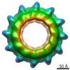
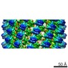
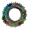
- PDB-3j9g: Atomic model of the VipA/VipB, the type six secretion system cont... -
+ Open data

- Basic information
| Entry | Database: PDB / ID: 3j9g | ||||||
|---|---|---|---|---|---|---|---|
| Title | Atomic model of the VipA/VipB, the type six secretion system contractile sheath of Vibrio cholerae from cryo-EM | ||||||
 Components Components |
| ||||||
 Keywords Keywords | CONTRACTILE PROTEIN / t6ss / bacterial secretion / phage / contraction | ||||||
| Function / homology | Type VI secretion system TssC-like / TssC1, N-terminal / TssC1, C-terminal / EvpB/VC_A0108, tail sheath N-terminal domain / EvpB/VC_A0108, tail sheath gpW/gp25-like domain / Type VI secretion system sheath protein TssB1 / Type VI secretion system, VipA, VC_A0107 or Hcp2 / Type VI secretion system contractile sheath large subunit / Type VI secretion system contractile sheath small subunit Function and homology information Function and homology information | ||||||
| Biological species |  Vibrio cholerae O1 biovar El Tor str. N16961 (bacteria) Vibrio cholerae O1 biovar El Tor str. N16961 (bacteria) | ||||||
| Method | ELECTRON MICROSCOPY / helical reconstruction / cryo EM / Resolution: 3.5 Å | ||||||
 Authors Authors | Kudryashev, M. / Wang, R.Y.-R. / Brackmann, M. / Scherer, S. / Maier, T. / Baker, D. / DiMaio, F. / Stahlberg, H. / Egelman, E.H. / Basler, M. | ||||||
 Citation Citation | Journal: Cell / Year: 2015 Title: Structure of the type VI secretion system contractile sheath. Authors: Mikhail Kudryashev / Ray Yu-Ruei Wang / Maximilian Brackmann / Sebastian Scherer / Timm Maier / David Baker / Frank DiMaio / Henning Stahlberg / Edward H Egelman / Marek Basler /   Abstract: Bacteria use rapid contraction of a long sheath of the type VI secretion system (T6SS) to deliver effectors into a target cell. Here, we present an atomic-resolution structure of a native contracted ...Bacteria use rapid contraction of a long sheath of the type VI secretion system (T6SS) to deliver effectors into a target cell. Here, we present an atomic-resolution structure of a native contracted Vibrio cholerae sheath determined by cryo-electron microscopy. The sheath subunits, composed of tightly interacting proteins VipA and VipB, assemble into a six-start helix. The helix is stabilized by a core domain assembled from four β strands donated by one VipA and two VipB molecules. The fold of inner and middle layers is conserved between T6SS and phage sheaths. However, the structure of the outer layer is distinct and suggests a mechanism of interaction of the bacterial sheath with an accessory ATPase, ClpV, that facilitates multiple rounds of effector delivery. Our results provide a mechanistic insight into assembly of contractile nanomachines that bacteria and phages use to translocate macromolecules across membranes. | ||||||
| History |
|
- Structure visualization
| Movie |
Movie viewer |
|---|---|
| Structure viewer | Molecule: MolmilJmol/JSmol |
- Downloads & links
-Download
| PDBx/mmCIF format | 3j9g.cif.gz | 3 MB | Display | PDBx/mmCIF format |
|---|---|---|---|---|
| PDB format | pdb3j9g.ent.gz | Display | PDB format | |
| PDBx/mmJSON format | 3j9g.json.gz | Tree view | PDBx/mmJSON format | |
| Others |  Other downloads Other downloads |
-Validation report
| Summary document | 3j9g_validation.pdf.gz | 1.3 MB | Display | wwPDB validaton report |
|---|---|---|---|---|
| Full document | 3j9g_full_validation.pdf.gz | 1.4 MB | Display | |
| Data in XML | 3j9g_validation.xml.gz | 355.5 KB | Display | |
| Data in CIF | 3j9g_validation.cif.gz | 556.1 KB | Display | |
| Arichive directory | https://data.pdbj.org/pub/pdb/validation_reports/j9/3j9gftp://data.pdbj.org/pub/pdb/validation_reports/j9/3j9g | HTTPS FTP |
-Related structure data
| Related structure data | 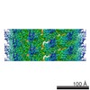 2699MC M: map data used to model this data C: citing same article ( |
|---|---|
| Similar structure data | |
| EM raw data | EMPIAR-10019 (Title: VipA/VipB, sheath of the bacterial type IV secretion system, micrographs for helical reconstruction taken on a K2 detector Data size: 4.1 Data #1: Frame-averaged micrographs [micrographs - single frame]) |

-Links
 PDBj
PDBj- Assembly
| Deposited unit | 
|
|---|---|
| 1 |
|
| Symmetry | Helical symmetry: (Circular symmetry: 6 / Dyad axis: no / N subunits divisor: 1 / Num. of operations: 5 / Rise per n subunits: 21.8 Å / Rotation per n subunits: 29.4 °) |
-Components
| #1: Protein | Mass: 13716.561 Da / Num. of mol.: 30 / Fragment: UNP residues 2-126 Source method: isolated from a genetically manipulated source Source: (gene. exp.) Vibrio cholerae O1 biovar El Tor str. N16961 (bacteria)Gene: VC_A0107 / Production host: Vibrio cholerae 2740-80 (bacteria) / References: UniProt: Q9KN58#2: Protein | Mass: 49167.539 Da / Num. of mol.: 30 / Fragment: UNP residues 61-492 Source method: isolated from a genetically manipulated source Source: (gene. exp.) Vibrio cholerae O1 biovar El Tor str. N16961 (bacteria)Gene: VC_A0108 / Production host: Vibrio cholerae 2740-80 (bacteria) / References: UniProt: Q9KN57 |
|---|
-Experimental details
-Experiment
| Experiment | Method: ELECTRON MICROSCOPY |
|---|---|
| EM experiment | Aggregation state: FILAMENT / 3D reconstruction method: helical reconstruction |
- Sample preparation
| Component |
| ||||||||||||||||
|---|---|---|---|---|---|---|---|---|---|---|---|---|---|---|---|---|---|
| Buffer solution | Name: PBS / pH: 7.4 / Details: PBS | ||||||||||||||||
| Specimen | Conc.: 0.1 mg/ml / Embedding applied: NO / Shadowing applied: NO / Staining applied: NO / Vitrification applied: YES | ||||||||||||||||
| Specimen support | Details: holey carbon grids, Quantifoil 1.2/1.3, imaged over the holes | ||||||||||||||||
| Vitrification | Instrument: FEI VITROBOT MARK IV / Cryogen name: ETHANE / Temp: 77 K / Details: Plunged into liquid ethane (FEI VITROBOT MARK IV) / Method: plunge freezing |
- Electron microscopy imaging
| Experimental equipment |  Model: Titan Krios / Image courtesy: FEI Company |
|---|---|
| Microscopy | Model: FEI TITAN KRIOS / Date: Nov 15, 2013 |
| Electron gun | Electron source:  FIELD EMISSION GUN / Accelerating voltage: 300 kV / Illumination mode: FLOOD BEAM FIELD EMISSION GUN / Accelerating voltage: 300 kV / Illumination mode: FLOOD BEAM |
| Electron lens | Mode: BRIGHT FIELD / Nominal magnification: 29000 X / Nominal defocus max: 2000 nm / Nominal defocus min: 400 nm / Cs: 2.7 mm |
| Specimen holder | Specimen holder model: FEI TITAN KRIOS AUTOGRID HOLDER / Temperature: 63 K / Tilt angle max: 5 ° / Tilt angle min: -5 ° |
| Image recording | Electron dose: 30 e/Å2 / Film or detector model: GATAN K2 (4k x 4k) / Details: Operated in counting non-superresolution mode |
| Image scans | Num. digital images: 72 |
- Processing
| EM software |
| |||||||||||||||
|---|---|---|---|---|---|---|---|---|---|---|---|---|---|---|---|---|
| CTF correction | Details: Phase Flip, CTF detection by CTFFIND | |||||||||||||||
| Helical symmerty | Angular rotation/subunit: 29.4 ° / Axial rise/subunit: 21.8 Å / Axial symmetry: C6 | |||||||||||||||
| 3D reconstruction | Method: Iterative Real Space Helical Reconstruction / Resolution: 3.5 Å / Num. of particles: 96000 / Nominal pixel size: 0.5 Å / Actual pixel size: 0.5 Å Magnification calibration: Magnification was calibrated in the microscope using a gold lattice. Num. of class averages: 1 / Symmetry type: HELICAL | |||||||||||||||
| Refinement step | Cycle: LAST
|
