Movie
Movie Controller

[English] 日本語
 Yorodumi
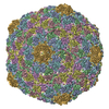
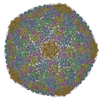
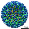
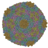
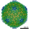
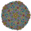
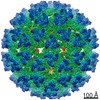
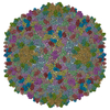
- PDB-3j1a: HK97-like fold fitted into 3D reconstruction of bacteriophage CW02 -
+ Open data

- Basic information
| Entry | Database: PDB / ID: 3j1a | ||||||
|---|---|---|---|---|---|---|---|
| Title | HK97-like fold fitted into 3D reconstruction of bacteriophage CW02 | ||||||
 Components Components | capsid protein | ||||||
 Keywords Keywords | VIRUS / halophage / bacteriophage HK97 / bacteriophage T7 / T7-like phage / turret / extremophile / Great Salt Lake | ||||||
| Function / homology | Phage capsid / Phage capsid family / viral procapsid maturation / T=7 icosahedral viral capsid / viral capsid / identical protein binding / Major capsid protein Function and homology information Function and homology information | ||||||
| Biological species | Great Salt Lake bacteriophage CW02 (others) | ||||||
| Method | ELECTRON MICROSCOPY / single particle reconstruction / cryo EM / Resolution: 16 Å | ||||||
 Authors Authors | Shen, P.S. / Domek, M.J. / Sanz-Garcia, E. / Makaju, A. / Taylor, R. / Culumber, M. / Breakwell, D.P. / Prince, J.T. / Belnap, D.M. | ||||||
 Citation Citation | Journal: J Virol / Year: 2012 Title: Sequence and structural characterization of great salt lake bacteriophage CW02, a member of the T7-like supergroup. Authors: Peter S Shen / Matthew J Domek / Eduardo Sanz-García / Aman Makaju / Ryan M Taylor / Ryan Hoggan / Michele D Culumber / Craig J Oberg / Donald P Breakwell / John T Prince / David M Belnap /  Abstract: Halophage CW02 infects a Salinivibrio costicola-like bacterium, SA50, isolated from the Great Salt Lake. Following isolation, cultivation, and purification, CW02 was characterized by DNA sequencing, ...Halophage CW02 infects a Salinivibrio costicola-like bacterium, SA50, isolated from the Great Salt Lake. Following isolation, cultivation, and purification, CW02 was characterized by DNA sequencing, mass spectrometry, and electron microscopy. A conserved module of structural genes places CW02 in the T7 supergroup, members of which are found in diverse aquatic environments, including marine and freshwater ecosystems. CW02 has morphological similarities to viruses of the Podoviridae family. The structure of CW02, solved by cryogenic electron microscopy and three-dimensional reconstruction, enabled the fitting of a portion of the bacteriophage HK97 capsid protein into CW02 capsid density, thereby providing additional evidence that capsid proteins of tailed double-stranded DNA phages have a conserved fold. The CW02 capsid consists of bacteriophage lambda gpD-like densities that likely contribute to particle stability. Turret-like densities were found on icosahedral vertices and may represent a unique adaptation similar to what has been seen in other extremophilic viruses that infect archaea, such as Sulfolobus turreted icosahedral virus and halophage SH1. | ||||||
| History |
|
- Structure visualization
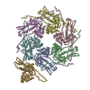
| Movie |
Movie viewer |
|---|---|
| Structure viewer | Molecule: MolmilJmol/JSmol |
- Downloads & links
-Download
| PDBx/mmCIF format | 3j1a.cif.gz | 49.9 KB | Display | PDBx/mmCIF format |
|---|---|---|---|---|
| PDB format | pdb3j1a.ent.gz | 28.3 KB | Display | PDB format |
| PDBx/mmJSON format | 3j1a.json.gz | Tree view | PDBx/mmJSON format | |
| Others |  Other downloads Other downloads |
-Validation report
| Summary document | 3j1a_validation.pdf.gz | 872.6 KB | Display | wwPDB validaton report |
|---|---|---|---|---|
| Full document | 3j1a_full_validation.pdf.gz | 872.2 KB | Display | |
| Data in XML | 3j1a_validation.xml.gz | 17.6 KB | Display | |
| Data in CIF | 3j1a_validation.cif.gz | 25.4 KB | Display | |
| Arichive directory | https://data.pdbj.org/pub/pdb/validation_reports/j1/3j1aftp://data.pdbj.org/pub/pdb/validation_reports/j1/3j1a | HTTPS FTP |
-Related structure data
| Related structure data | 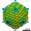 5388MC M: map data used to model this data C: citing same article ( |
|---|---|
| Similar structure data |
-Links
 PDBj
PDBj

- Assembly
| Deposited unit | 
|
|---|---|
| 1 | x 60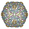
|
| 2 |
|
| 3 | x 5
|
| 4 | x 6
|
| 5 | 
|
| Symmetry | Point symmetry: (Schoenflies symbol: I (icosahedral)) |
-Components
| #1: Protein | Mass: 21944.676 Da / Num. of mol.: 7 / Fragment: SEE REMARK 999 / Source method: isolated from a natural source Source: (natural) Great Salt Lake bacteriophage CW02 (others) References: UniProt: P49861*PLUS Sequence details | MODELED SEQUENCE COMPRISES RESIDUES 182-380 OF MAJOR CAPSID PROTEIN FROM ENTEROBACTERIA PHAGE HK97 ...MODELED SEQUENCE COMPRISES RESIDUES 182-380 OF MAJOR CAPSID PROTEIN FROM ENTEROBACT | |
|---|
-Experimental details
-Experiment
| Experiment | Method: ELECTRON MICROSCOPY |
|---|---|
| EM experiment | Aggregation state: PARTICLE / 3D reconstruction method: single particle reconstruction |
- Sample preparation
| Component | Name: Bacteriophage CW02 / Type: VIRUS Details: Head and tail phage. Sample buffer solution contains 8% NaCl |
|---|---|
| Details of virus | Empty: NO / Enveloped: NO / Host category: BACTERIA(EUBACTERIA) / Isolate: SPECIES / Type: VIRION |
| Natural host | Organism: Salinivibrio costicola |
| Buffer solution | pH: 8 Details: 1.35 M NaCl, 48 mM MgSO4-7H2O, 1 mM CaCl2, 2 mM Tris-Cl |
| Specimen | Conc.: 3 mg/ml / Embedding applied: NO / Shadowing applied: NO / Staining applied: NO / Vitrification applied: YES Details: 1.35 M NaCl, 48 mM MgSO4-7H2O, 1 mM CaCl2, 2 mM Tris-Cl |
| Specimen support | Details: 200 mesh, holey-carbon-coated copper grid |
| Vitrification | Instrument: FEI VITROBOT MARK III / Cryogen name: ETHANE / Humidity: 100 % / Chamber temperature: 277 K Details: Blotted for 4 seconds before plunging into liquid ethane (FEI Vitrobot MarK III) Method: Blot for 4 seconds before plunging |
- Electron microscopy imaging
| Experimental equipment |  Model: Tecnai F30 / Image courtesy: FEI Company |
|---|---|
| Microscopy | Model: FEI TECNAI F30 / Date: Jul 1, 2009 |
| Electron gun | Electron source:  FIELD EMISSION GUN / Accelerating voltage: 200 kV / Illumination mode: FLOOD BEAM FIELD EMISSION GUN / Accelerating voltage: 200 kV / Illumination mode: FLOOD BEAM |
| Electron lens | Mode: BRIGHT FIELD / Nominal magnification: 39000 X / Calibrated magnification: 37564 X / Nominal defocus max: 2800 nm / Nominal defocus min: 200 nm / Cs: 2 mm / Camera length: 0 mm |
| Specimen holder | Specimen holder model: GATAN LIQUID NITROGEN / Temperature: 93 K / Temperature (max): 94 K / Temperature (min): 92 K / Tilt angle max: 0 ° / Tilt angle min: 0 ° |
| Image recording | Film or detector model: KODAK SO-163 FILM |
| Radiation | Protocol: SINGLE WAVELENGTH / Monochromatic (M) / Laue (L): M / Scattering type: x-ray |
| Radiation wavelength | Relative weight: 1 |
- Processing
| EM software |
| ||||||||||||||||||||
|---|---|---|---|---|---|---|---|---|---|---|---|---|---|---|---|---|---|---|---|---|---|
| CTF correction | Details: whole micrograph | ||||||||||||||||||||
| Symmetry | Point symmetry: I (icosahedral) | ||||||||||||||||||||
| 3D reconstruction | Method: projection matching / Resolution: 16 Å / Resolution method: FSC 0.5 CUT-OFF / Num. of particles: 8695 / Details: Particles were manually selected using X3D / Symmetry type: POINT | ||||||||||||||||||||
| Atomic model building | Protocol: RIGID BODY FIT / Space: REAL Details: REFINEMENT PROTOCOL--Automatic rigid body DETAILS--C-alpha coordinates pertaining to the HK97-fold were separately fitted as rigid bodies into capsid hexamers or pentamers. | ||||||||||||||||||||
| Atomic model building | PDB-ID: 1OHG Pdb chain-ID: G / Accession code: 1OHG / Source name: PDB / Type: experimental model | ||||||||||||||||||||
| Refinement step | Cycle: LAST
|