Movie
Movie Controller

[English] 日本語
 Yorodumi
- EMDB-5388: Cryo-electron microscopy and three-dimensional reconstruction of ... -
+ Open data

- Basic information
| Entry | Database: EMDB / ID: EMD-5388 | |||||||||
|---|---|---|---|---|---|---|---|---|---|---|
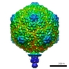
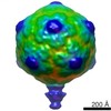
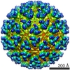
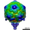
| Title | Cryo-electron microscopy and three-dimensional reconstruction of bacteriophage CW02 | |||||||||
 Map data Map data | EMD-5388 | |||||||||
 Sample Sample |
| |||||||||
 Keywords Keywords | halophage /  bacteriophage / bacteriophage HK97 / bacteriophage T7 / T7-like phage / turret / extremophile / Great Salt Lake bacteriophage / bacteriophage HK97 / bacteriophage T7 / T7-like phage / turret / extremophile / Great Salt Lake | |||||||||
| Function / homology | Phage capsid / Phage capsid family / viral procapsid maturation / T=7 icosahedral viral capsid / viral capsid / identical protein binding / Major capsid protein Function and homology information Function and homology information | |||||||||
| Biological species |  Bacteriophage CW02 (virus) Bacteriophage CW02 (virus) | |||||||||
| Method | single particle reconstruction / cryo EM / Resolution: 16.0 Å | |||||||||
 Authors Authors | Shen PS / Domek MJ / Sanz-Garcia E / Makaju A / Taylor R / Culumber M / Breakwell DP / Prince JT / Belnap DM | |||||||||
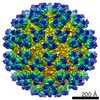
 Citation Citation | Journal: J Virol / Year: 2012 Title: Sequence and structural characterization of great salt lake bacteriophage CW02, a member of the T7-like supergroup. Authors: Peter S Shen / Matthew J Domek / Eduardo Sanz-García / Aman Makaju / Ryan M Taylor / Ryan Hoggan / Michele D Culumber / Craig J Oberg / Donald P Breakwell / John T Prince / David M Belnap /  Abstract: Halophage CW02 infects a Salinivibrio costicola-like bacterium, SA50, isolated from the Great Salt Lake. Following isolation, cultivation, and purification, CW02 was characterized by DNA sequencing, ...Halophage CW02 infects a Salinivibrio costicola-like bacterium, SA50, isolated from the Great Salt Lake. Following isolation, cultivation, and purification, CW02 was characterized by DNA sequencing, mass spectrometry, and electron microscopy. A conserved module of structural genes places CW02 in the T7 supergroup, members of which are found in diverse aquatic environments, including marine and freshwater ecosystems. CW02 has morphological similarities to viruses of the Podoviridae family. The structure of CW02, solved by cryogenic electron microscopy and three-dimensional reconstruction, enabled the fitting of a portion of the bacteriophage HK97 capsid protein into CW02 capsid density, thereby providing additional evidence that capsid proteins of tailed double-stranded DNA phages have a conserved fold. The CW02 capsid consists of bacteriophage lambda gpD-like densities that likely contribute to particle stability. Turret-like densities were found on icosahedral vertices and may represent a unique adaptation similar to what has been seen in other extremophilic viruses that infect archaea, such as Sulfolobus turreted icosahedral virus and halophage SH1. | |||||||||
| History |
|
- Structure visualization
| Movie |
Movie viewer |
|---|---|
| Structure viewer | EM map: SurfViewMolmilJmol/JSmol |
| Supplemental images |

- Downloads & links
-EMDB archive
| Map data | emd_5388.map.gz | 34.8 MB | EMDB map data format | |
|---|---|---|---|---|
| Header (meta data) | emd-5388-v30.xmlemd-5388.xml | 11.8 KB 11.8 KB | Display Display | EMDB header |
| Images | 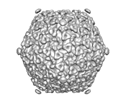 emd_5388_1.jpg emd_5388_1.jpg | 42.9 KB | ||
| Archive directory |  http://ftp.pdbj.org/pub/emdb/structures/EMD-5388ftp://ftp.pdbj.org/pub/emdb/structures/EMD-5388 http://ftp.pdbj.org/pub/emdb/structures/EMD-5388ftp://ftp.pdbj.org/pub/emdb/structures/EMD-5388 | HTTPS FTP |
-Related structure data
| Related structure data |  3j1aMC M: atomic model generated by this map C: citing same article ( |
|---|---|
| Similar structure data |

-Links
| EMDB pages | EMDB (EBI/PDBe) / EMDataResource |
|---|---|
| Related items in Molecule of the Month |


-Map
| File | Download / File: emd_5388.map.gz / Format: CCP4 / Size: 68.5 MB / Type: IMAGE STORED AS FLOATING POINT NUMBER (4 BYTES) | ||||||||||||||||||||||||||||||||||||||||||||||||||||||||||||||||||||
|---|---|---|---|---|---|---|---|---|---|---|---|---|---|---|---|---|---|---|---|---|---|---|---|---|---|---|---|---|---|---|---|---|---|---|---|---|---|---|---|---|---|---|---|---|---|---|---|---|---|---|---|---|---|---|---|---|---|---|---|---|---|---|---|---|---|---|---|---|---|
| Annotation | EMD-5388 | ||||||||||||||||||||||||||||||||||||||||||||||||||||||||||||||||||||
| Voxel size | X=Y=Z: 3.14 Å | ||||||||||||||||||||||||||||||||||||||||||||||||||||||||||||||||||||
| Density |
| ||||||||||||||||||||||||||||||||||||||||||||||||||||||||||||||||||||
| Symmetry | Space group: 1 | ||||||||||||||||||||||||||||||||||||||||||||||||||||||||||||||||||||
| Details | EMDB XML:
CCP4 map header:
| ||||||||||||||||||||||||||||||||||||||||||||||||||||||||||||||||||||
-Supplemental data
- Sample components
-Entire : Bacteriophage CW02
| Entire | Name: Bacteriophage CW02 (virus) |
|---|---|
| Components |
|
-Supramolecule #1000: Bacteriophage CW02
| Supramolecule | Name: Bacteriophage CW02 / type: sample / ID: 1000 / Details: Sample buffer solution contains 8% NaCl / Oligomeric state: head and tail phage / Number unique components: 1 |
|---|
-Supramolecule #1: Bacteriophage CW02
| Supramolecule | Name: Bacteriophage CW02 / type: virus / ID: 1 / Sci species name: Bacteriophage CW02 / Database: NCBI / Virus type: VIRION / Virus isolate: SPECIES / Virus enveloped: No / Virus empty: No |
|---|---|
| Host (natural) | Organism:  Salinivibrio costicola (bacteria) / synonym: BACTERIA(EUBACTERIA) Salinivibrio costicola (bacteria) / synonym: BACTERIA(EUBACTERIA) |
| Virus shell | Shell ID: 1 / Name: gp48 / Diameter: 691 Å / T number (triangulation number): 7 |
-Experimental details
-Structure determination
| Method | cryo EM |
|---|---|
 Processing Processing | single particle reconstruction |
| Aggregation state | particle |
-Sample preparation
| Concentration | 3 mg/mL |
|---|---|
| Buffer | pH: 8 Details: 1.35 M NaCl, 48 mM MgSO4-7H2O, 1 mM CaCl2, 2 mM Tris-Cl |
| Grid | Details: 200 mesh, holey-carbon-coated copper grid |
| Vitrification | Cryogen name: ETHANE / Chamber humidity: 100 % / Chamber temperature: 277 K / Instrument: FEI VITROBOT MARK III / Method: Blot for 4 seconds before plunging |
- Electron microscopy
| Microscope | FEI TECNAI F30 |
|---|---|
| Electron beam | Acceleration voltage: 200 kV / Electron source: FIELD EMISSION GUN |
| Electron optics | Calibrated magnification: 37564 / Illumination mode: FLOOD BEAM / Imaging mode: BRIGHT FIELDBright-field microscopy / Cs: 2.0 mm / Nominal defocus max: 2.8 µm / Nominal defocus min: 0.2 µm / Nominal magnification: 39000 |
| Sample stage | Specimen holder model: GATAN LIQUID NITROGEN |
| Temperature | Min: 92 K / Max: 94 K / Average: 93 K |
| Date | Jul 1, 2009 |
| Image recording | Category: FILM / Film or detector model: KODAK SO-163 FILM / Digitization - Scanner: NIKON SUPER COOLSCAN 9000 / Digitization - Sampling interval: 1.57 µm / Number real images: 8695 |
| Experimental equipment |  Model: Tecnai F30 / Image courtesy: FEI Company |
-Image processing
| CTF correction | Details: whole micrograph |
|---|---|
| Final angle assignment | Details: PFT2 |
| Final reconstruction | Algorithm: OTHER / Resolution.type: BY AUTHOR / Resolution: 16.0 Å / Resolution method: FSC 0.5 CUT-OFF / Software - Name: PFT2,EM3DR2,(PFT3DR) / Number images used: 8695 |
| Details | Particles were manually selected using X3D |
-Atomic model buiding 1
| Initial model | PDB ID: Chain - #0 - Chain ID: A / Chain - #1 - Chain ID: B / Chain - #2 - Chain ID: C / Chain - #3 - Chain ID: D / Chain - #4 - Chain ID: E / Chain - #5 - Chain ID: F / Chain - #6 - Chain ID: G |
|---|---|
| Software | Name: Chimera |
| Details | Protocol: Automatic rigid body. C-alpha coordinates pertaining to the HK97-fold were separately fitted as rigid bodies into capsid hexamers or pentamers. |
| Refinement | Space: REAL / Protocol: RIGID BODY FIT |
| Output model | PDB-3j1a: |