 Movie
Movie Controller

[English] 日本語
 Yorodumi
- EMDB-5448: Cryo-tomography and subtomogram averaging of the Newcastle diseas... -
+ Open data

- Basic information
| Entry | Database: EMDB / ID: EMD-5448 | |||||||||
|---|---|---|---|---|---|---|---|---|---|---|
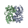
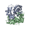
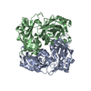
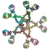
| Title | Cryo-tomography and subtomogram averaging of the Newcastle disease virus matrix protein array | |||||||||
 Map data Map data | Subtomogram average of Newcastle disease virus matrix protein | |||||||||
 Sample Sample |
| |||||||||
 Keywords Keywords | virus assembly / matrix protein / pleomorphic virus structure / paramyxovirus / viral membrane / cryo-tomography | |||||||||
| Biological species |  Newcastle disease virus Newcastle disease virus | |||||||||
| Method | subtomogram averaging / cryo EM / Resolution: 45.6 Å | |||||||||
 Authors Authors | Battisti AJ / Meng G / Winkler DC / McGinnes LW / Plevka P / Steven AC / Morrison TG / Rossmann MG | |||||||||
 Citation Citation | Journal: Proc Natl Acad Sci U S A / Year: 2012 Title: Structure and assembly of a paramyxovirus matrix protein. Authors: Anthony J Battisti / Geng Meng / Dennis C Winkler / Lori W McGinnes / Pavel Plevka / Alasdair C Steven / Trudy G Morrison / Michael G Rossmann /  Abstract: Many pleomorphic, lipid-enveloped viruses encode matrix proteins that direct their assembly and budding, but the mechanism of this process is unclear. We have combined X-ray crystallography and ...Many pleomorphic, lipid-enveloped viruses encode matrix proteins that direct their assembly and budding, but the mechanism of this process is unclear. We have combined X-ray crystallography and cryoelectron tomography to show that the matrix protein of Newcastle disease virus, a paramyxovirus and relative of measles virus, forms dimers that assemble into pseudotetrameric arrays that generate the membrane curvature necessary for virus budding. We show that the glycoproteins are anchored in the gaps between the matrix proteins and that the helical nucleocapsids are associated in register with the matrix arrays. About 90% of virions lack matrix arrays, suggesting that, in agreement with previous biological observations, the matrix protein needs to dissociate from the viral membrane during maturation, as is required for fusion and release of the nucleocapsid into the host's cytoplasm. Structure and sequence conservation imply that other paramyxovirus matrix proteins function similarly. | |||||||||
| History |
|
- Structure visualization
| Movie |
 Movie viewer Movie viewer |
|---|---|
| Structure viewer | EM map: SurfViewMolmilJmol/JSmol |
| Supplemental images |

- Downloads & links
-EMDB archive
| Map data | emd_5448.map.gz | 169.8 KB | EMDB map data format | |
|---|---|---|---|---|
| Header (meta data) | emd-5448-v30.xmlemd-5448.xml | 11.4 KB 11.4 KB | Display Display | EMDB header |
| Images | emd_5448.tif | 161 KB | ||
| Archive directory |  http://ftp.pdbj.org/pub/emdb/structures/EMD-5448ftp://ftp.pdbj.org/pub/emdb/structures/EMD-5448 http://ftp.pdbj.org/pub/emdb/structures/EMD-5448ftp://ftp.pdbj.org/pub/emdb/structures/EMD-5448 | HTTPS FTP |
-Validation report
| Summary document | emd_5448_validation.pdf.gz | 78.3 KB | Display | EMDB validaton report |
|---|---|---|---|---|
| Full document | emd_5448_full_validation.pdf.gz | 77.4 KB | Display | |
| Data in XML | emd_5448_validation.xml.gz | 495 B | Display | |
| Arichive directory | https://ftp.pdbj.org/pub/emdb/validation_reports/EMD-5448ftp://ftp.pdbj.org/pub/emdb/validation_reports/EMD-5448 | HTTPS FTP |
-Related structure data
-Links
| EMDB pages | EMDB (EBI/PDBe) / EMDataResource |
|---|
-Map
| File | Download / File: emd_5448.map.gz / Format: CCP4 / Size: 1 MB / Type: IMAGE STORED AS FLOATING POINT NUMBER (4 BYTES) | ||||||||||||||||||||||||||||||||||||||||||||||||||||||||||||||||||||
|---|---|---|---|---|---|---|---|---|---|---|---|---|---|---|---|---|---|---|---|---|---|---|---|---|---|---|---|---|---|---|---|---|---|---|---|---|---|---|---|---|---|---|---|---|---|---|---|---|---|---|---|---|---|---|---|---|---|---|---|---|---|---|---|---|---|---|---|---|---|
| Annotation | Subtomogram average of Newcastle disease virus matrix protein | ||||||||||||||||||||||||||||||||||||||||||||||||||||||||||||||||||||
| Voxel size | X=Y=Z: 7.5 Å | ||||||||||||||||||||||||||||||||||||||||||||||||||||||||||||||||||||
| Density |
| ||||||||||||||||||||||||||||||||||||||||||||||||||||||||||||||||||||
| Symmetry | Space group: 1 | ||||||||||||||||||||||||||||||||||||||||||||||||||||||||||||||||||||
| Details | EMDB XML:
CCP4 map header:
| ||||||||||||||||||||||||||||||||||||||||||||||||||||||||||||||||||||
-Supplemental data
- Sample components
-Entire : Newcastle disease virus matrix protein array from subtomogram ave...
| Entire | Name: Newcastle disease virus matrix protein array from subtomogram averaging |
|---|---|
| Components |
|
-Supramolecule #1000: Newcastle disease virus matrix protein array from subtomogram ave...
| Supramolecule | Name: Newcastle disease virus matrix protein array from subtomogram averaging type: sample / ID: 1000 / Oligomeric state: an array of dimers / Number unique components: 1 |
|---|---|
| Molecular weight | Theoretical: 76 KDa |
-Supramolecule #1: Newcastle disease virus
| Supramolecule | Name: Newcastle disease virus / type: virus / ID: 1 / Details: Newcastle disease virus strain B1 / NCBI-ID: 11176 / Sci species name: Newcastle disease virus / Database: NCBI / Virus type: VIRION / Virus isolate: STRAIN / Virus enveloped: Yes / Virus empty: No |
|---|---|
| Host (natural) | Organism:  |
-Experimental details
-Structure determination
| Method | cryo EM |
|---|---|
 Processing Processing | subtomogram averaging |
-Sample preparation
| Buffer | pH: 8 / Details: 0.02 M Tris, 0.12 M NaCl, 0.001 M EDTA |
|---|---|
| Grid | Details: 200 mesh holey carbon copper grids (Quantifoil Micro Tools, GmbH, Germany) |
| Vitrification | Cryogen name: ETHANE / Instrument: HOMEMADE PLUNGER |
- Electron microscopy
| Microscope | FEI TITAN KRIOS |
|---|---|
| Alignment procedure | Legacy - Astigmatism: Objective lens astigmatism was corrected at 19,500 times magnification |
| Specialist optics | Energy filter - Name: Tridiem GIF 863 / Energy filter - Lower energy threshold: 0.0 eV / Energy filter - Upper energy threshold: 30.0 eV |
| Date | Oct 19, 2010 |
| Image recording | Category: CCD / Film or detector model: GATAN ULTRASCAN 1000 (2k x 2k) / Digitization - Sampling interval: 15.0 µm / Number real images: 87 / Average electron dose: 163 e/Å2 / Bits/pixel: 16 |
| Electron beam | Acceleration voltage: 300 kV / Electron source:  FIELD EMISSION GUN FIELD EMISSION GUN |
| Electron optics | Calibrated magnification: 20000 / Illumination mode: FLOOD BEAM / Imaging mode: BRIGHT FIELD / Cs: 2.7 mm / Nominal defocus max: 8.0 µm / Nominal defocus min: 8.0 µm / Nominal magnification: 19500 |
| Sample stage | Specimen holder model: OTHER / Tilt series - Axis1 - Min angle: -64.5 ° / Tilt series - Axis1 - Max angle: 64.5 ° |
| Experimental equipment |  Model: Titan Krios / Image courtesy: FEI Company |
-Image processing
| Details | Images aligned using colloidal gold fiducial markers and reconstructed using the weighted back-projection method as implemented in EMAN. Average number of tilts used in the 3D reconstructions: 87. Average tomographic tilt angle increment: 1.5. |
|---|---|
| Final reconstruction | Algorithm: OTHER / Resolution.type: BY AUTHOR / Resolution: 45.6 Å / Resolution method: FSC 0.5 CUT-OFF / Software - Name: IMOD, Uppsala_Software_Factory Details: Tomographic reconstruction showed an array of matrix protein subunits, which were averaged to reduce noise. Two-fold symmetry enforced for individual subunits. Membrane density masked out. |
-Atomic model buiding 1
| Initial model | PDB ID: Chain - #0 - Chain ID: A / Chain - #1 - Chain ID: B |
|---|---|
| Software | Name: EMfit |
| Details | Protocol: Rigid body. 10 matrix protein dimers were simultaneously fitted into the matrix array density using EMfit |
| Refinement | Space: REAL / Protocol: RIGID BODY FIT / Target criteria: sumF, clash |
