 Movie
Movie Controller

[English] 日本語
 Yorodumi
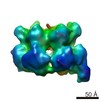
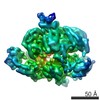
- EMDB-2872: Negative stain electron microscopy of recombinant human minichrom... -
+ Open data

- Basic information
| Entry | Database: EMDB / ID: EMD-2872 | |||||||||
|---|---|---|---|---|---|---|---|---|---|---|
| Title | Negative stain electron microscopy of recombinant human minichromosome maintenance complex | |||||||||
 Map data Map data | Reconstruction of recombinant human minichromosome maintenance complex | |||||||||
 Sample Sample |
| |||||||||
 Keywords Keywords | mini chromosome maintenance / DNA helicase / MCM2-7 / hMCM | |||||||||
| Biological species |  Homo sapiens (human) Homo sapiens (human) | |||||||||
| Method | single particle reconstruction / negative staining / Resolution: 23.0 Å | |||||||||
 Authors Authors | Hesketh EL / Parker-Manuel RP / Chaban Y / Satti R / Coverley D / Orlova EV / Chong JPJ | |||||||||
 Citation Citation | Journal: J Biol Chem / Year: 2015 Title: DNA induces conformational changes in a recombinant human minichromosome maintenance complex. Authors: Emma L Hesketh / Richard P Parker-Manuel / Yuriy Chaban / Rabab Satti / Dawn Coverley / Elena V Orlova / James P J Chong /  Abstract: ATP-dependent DNA unwinding activity has been demonstrated for recombinant archaeal homohexameric minichromosome maintenance (MCM) complexes and their yeast heterohexameric counterparts, but in ...ATP-dependent DNA unwinding activity has been demonstrated for recombinant archaeal homohexameric minichromosome maintenance (MCM) complexes and their yeast heterohexameric counterparts, but in higher eukaryotes such as Drosophila, MCM-associated DNA helicase activity has been observed only in the context of a co-purified Cdc45-MCM-GINS complex. Here, we describe the production of the recombinant human MCM (hMCM) complex in Escherichia coli. This protein displays ATP hydrolysis activity and is capable of unwinding duplex DNA. Using single-particle asymmetric EM reconstruction, we demonstrate that recombinant hMCM forms a hexamer that undergoes a conformational change when bound to DNA. Recombinant hMCM produced without post-translational modifications is functional in vitro and provides an important tool for biochemical reconstitution of the human replicative helicase. | |||||||||
| History |
|
- Structure visualization
| Movie |
 Movie viewer Movie viewer |
|---|---|
| Structure viewer | EM map: SurfViewMolmilJmol/JSmol |
| Supplemental images |
 UCSF Chimera
- Downloads & links
-EMDB archive
| Map data | emd_2872.map.gz | 1.6 MB | EMDB map data format | |
|---|---|---|---|---|
| Header (meta data) | emd-2872-v30.xmlemd-2872.xml | 9 KB 9 KB | Display Display | EMDB header |
| Images | 3.tif | 160.4 KB | ||
| Archive directory |  http://ftp.pdbj.org/pub/emdb/structures/EMD-2872ftp://ftp.pdbj.org/pub/emdb/structures/EMD-2872 http://ftp.pdbj.org/pub/emdb/structures/EMD-2872ftp://ftp.pdbj.org/pub/emdb/structures/EMD-2872 | HTTPS FTP |
-Validation report
| Summary document | emd_2872_validation.pdf.gz | 180.5 KB | Display | EMDB validaton report |
|---|---|---|---|---|
| Full document | emd_2872_full_validation.pdf.gz | 179.7 KB | Display | |
| Data in XML | emd_2872_validation.xml.gz | 5.3 KB | Display | |
| Arichive directory | https://ftp.pdbj.org/pub/emdb/validation_reports/EMD-2872ftp://ftp.pdbj.org/pub/emdb/validation_reports/EMD-2872 | HTTPS FTP |
-Related structure data


-Links
| EMDB pages | EMDB (EBI/PDBe) / EMDataResource |
|---|
-Map
| File | Download / File: emd_2872.map.gz / Format: CCP4 / Size: 29.8 MB / Type: IMAGE STORED AS FLOATING POINT NUMBER (4 BYTES) | ||||||||||||||||||||||||||||||||||||||||||||||||||||||||||||
|---|---|---|---|---|---|---|---|---|---|---|---|---|---|---|---|---|---|---|---|---|---|---|---|---|---|---|---|---|---|---|---|---|---|---|---|---|---|---|---|---|---|---|---|---|---|---|---|---|---|---|---|---|---|---|---|---|---|---|---|---|---|
| Annotation | Reconstruction of recombinant human minichromosome maintenance complex | ||||||||||||||||||||||||||||||||||||||||||||||||||||||||||||
| Voxel size | X=Y=Z: 2.5 Å | ||||||||||||||||||||||||||||||||||||||||||||||||||||||||||||
| Density |
| ||||||||||||||||||||||||||||||||||||||||||||||||||||||||||||
| Symmetry | Space group: 1 | ||||||||||||||||||||||||||||||||||||||||||||||||||||||||||||
| Details | EMDB XML:
CCP4 map header:
| ||||||||||||||||||||||||||||||||||||||||||||||||||||||||||||
-Supplemental data
- Sample components
-Entire : Recombinant human mini chromosome maintenance complex 2-7
| Entire | Name: Recombinant human mini chromosome maintenance complex 2-7 |
|---|---|
| Components |
|
-Supramolecule #1000: Recombinant human mini chromosome maintenance complex 2-7
| Supramolecule | Name: Recombinant human mini chromosome maintenance complex 2-7 type: sample / ID: 1000 / Number unique components: 1 |
|---|---|
| Molecular weight | Theoretical: 567 KDa |
-Macromolecule #1: Mini chromosome maintenance (2-7) complex
| Macromolecule | Name: Mini chromosome maintenance (2-7) complex / type: protein_or_peptide / ID: 1 / Name.synonym: MCM, hMCM / Number of copies: 1 / Oligomeric state: heterohexamer / Recombinant expression: Yes |
|---|---|
| Source (natural) | Organism: Homo sapiens (human) / synonym: Human |
| Molecular weight | Theoretical: 567 KDa |
| Recombinant expression | Organism:  |
-Experimental details
-Structure determination
| Method | negative staining |
|---|---|
 Processing Processing | single particle reconstruction |
| Aggregation state | particle |
-Sample preparation
| Concentration | 0.999 mg/mL |
|---|---|
| Buffer | pH: 8 Details: 25 mM HEPES pH 8.0, 200mM sodium glutamate, 1mM DTT, 0.1 mM AEBSF |
| Staining | Type: NEGATIVE / Details: Stained with methylamine tungstate |
| Grid | Details: Carbon coated grids |
| Vitrification | Cryogen name: NONE / Instrument: OTHER |
- Electron microscopy
| Microscope | FEI TECNAI 12 |
|---|---|
| Date | May 10, 2012 |
| Image recording | Category: FILM / Film or detector model: KODAK SO-163 FILM / Digitization - Scanner: ZEISS SCAI / Digitization - Sampling interval: 14 µm / Number real images: 31 |
| Electron beam | Acceleration voltage: 120 kV / Electron source: LAB6 |
| Electron optics | Calibrated magnification: 67000 / Illumination mode: SPOT SCAN / Imaging mode: DARK FIELD |
| Sample stage | Specimen holder model: SIDE ENTRY, EUCENTRIC |
-Image processing
| Details | Particle picking was carried out automatically using the program Boxer EMAN suite (Tang et al., 2007). Analysis of the CTF and correction was completed using the program CTFIT (EMAN suite, Tang et al., 2007). The following image analysis was performed using IMAGIC-5 (van Heel et al., 1996). |
|---|---|
| CTF correction | Details: EACH MICROGRAPH |
| Final reconstruction | Applied symmetry - Point group: C1 (asymmetric) / Algorithm: OTHER / Resolution.type: BY AUTHOR / Resolution: 23.0 Å / Resolution method: OTHER / Software - Name: IMAGIC / Details: Final maps were averaged from 10 classes / Number images used: 1100 |
| Final two d classification | Number classes: 100 |
