 Movie
Movie Controller

[English] 日本語
 Yorodumi
- PDB-7cg3: Staggered ring conformation of CtHsp104 (Hsp104 from Chaetomium T... -
+ Open data

- Basic information
| Entry | Database: PDB / ID: 7cg3 | |||||||||||||||
|---|---|---|---|---|---|---|---|---|---|---|---|---|---|---|---|---|
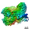
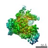
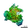
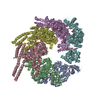
| Title | Staggered ring conformation of CtHsp104 (Hsp104 from Chaetomium Thermophilum) | |||||||||||||||
 Components Components | Heat shock protein 104 Heat shock response Heat shock response | |||||||||||||||
 Keywords Keywords | CHAPERONE / AAA+ ATPase / disaggregation | |||||||||||||||
| Function / homology |  Function and homology information Function and homology information | |||||||||||||||
| Biological species |  Chaetomium thermophilum var. coprophilum (fungus) Chaetomium thermophilum var. coprophilum (fungus) | |||||||||||||||
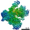
| Method | ELECTRON MICROSCOPY / single particle reconstruction / cryo EM / Resolution: 5.1 Å | |||||||||||||||
 Authors Authors | Inoue, Y. / Hanazono, Y. / Noi, K. / Kawamoto, A. / Kimatsuka, M. / Harada, R. / Takeda, K. / Iwamasa, N. / Shibata, K. / Noguchi, K. ...Inoue, Y. / Hanazono, Y. / Noi, K. / Kawamoto, A. / Kimatsuka, M. / Harada, R. / Takeda, K. / Iwamasa, N. / Shibata, K. / Noguchi, K. / Shigeta, Y. / Namba, K. / Ogura, T. / Miki, K. / Shinohara, K. / Yohda, M. | |||||||||||||||
| Funding support |  Japan, 4items Japan, 4items
| |||||||||||||||
 Citation Citation | Journal: Structure / Year: 2021 Title: Split conformation of Chaetomium thermophilum Hsp104 disaggregase. Authors: Yosuke Inoue / Yuya Hanazono / Kentaro Noi / Akihiro Kawamoto / Masato Kimatsuka / Ryuhei Harada / Kazuki Takeda / Ryoichi Kita / Natsuki Iwamasa / Kyoka Shibata / Keiichi Noguchi / Yasuteru ...Authors: Yosuke Inoue / Yuya Hanazono / Kentaro Noi / Akihiro Kawamoto / Masato Kimatsuka / Ryuhei Harada / Kazuki Takeda / Ryoichi Kita / Natsuki Iwamasa / Kyoka Shibata / Keiichi Noguchi / Yasuteru Shigeta / Keiichi Namba / Teru Ogura / Kunio Miki / Kyosuke Shinohara / Masafumi Yohda / Abstract: Hsp104 and its bacterial homolog ClpB form hexameric ring structures and mediate protein disaggregation. The disaggregated polypeptide is thought to thread through the central channel of the ring. ...Hsp104 and its bacterial homolog ClpB form hexameric ring structures and mediate protein disaggregation. The disaggregated polypeptide is thought to thread through the central channel of the ring. However, the dynamic behavior of Hsp104 during disaggregation remains unclear. Here, we reported the stochastic conformational dynamics and a split conformation of Hsp104 disaggregase from Chaetomium thermophilum (CtHsp104) in the presence of ADP by X-ray crystallography, cryo-electron microscopy (EM), and high-speed atomic force microscopy (AFM). ADP-bound CtHsp104 assembles into a 6 left-handed spiral filament in the crystal structure at a resolution of 2.7 Å. The unit of the filament is a hexamer of the split spiral structure. In the cryo-EM images, staggered and split hexameric rings were observed. Further, high-speed AFM observations showed that a substrate addition enhanced the conformational change and increased the split structure's frequency. Our data suggest that split conformation is an off-pathway state of CtHsp104 during disaggregation. | |||||||||||||||
| History |
|
- Structure visualization
| Movie |
Movie viewer |
|---|---|
| Structure viewer | Molecule: MolmilJmol/JSmol |
- Downloads & links
-Download
| PDBx/mmCIF format | 7cg3.cif.gz | 635.2 KB | Display | PDBx/mmCIF format |
|---|---|---|---|---|
| PDB format | pdb7cg3.ent.gz | 541.3 KB | Display | PDB format |
| PDBx/mmJSON format | 7cg3.json.gz | Tree view | PDBx/mmJSON format | |
| Others |  Other downloads Other downloads |
-Validation report
| Arichive directory | https://data.pdbj.org/pub/pdb/validation_reports/cg/7cg3ftp://data.pdbj.org/pub/pdb/validation_reports/cg/7cg3 | HTTPS FTP |
|---|
-Related structure data
| Related structure data | 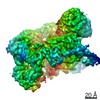 30349MC  5zuiC M: map data used to model this data C: citing same article ( |
|---|---|
| Similar structure data |


-Links
 PDBj
PDBj


- Assembly
| Deposited unit | 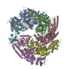
|
|---|---|
| 1 |
|
-Components
| #1: Protein | Heat shock response Mass: 85056.070 Da / Num. of mol.: 6 Source method: isolated from a genetically manipulated source Source: (gene. exp.) Chaetomium thermophilum var. coprophilum (fungus)Gene: hsp104 / Production host:  Escherichia coli BL21(DE3) (bacteria) / Strain (production host): BL21(DE3) / References: UniProt: A0A2Z6G185 Escherichia coli BL21(DE3) (bacteria) / Strain (production host): BL21(DE3) / References: UniProt: A0A2Z6G185 |
|---|
-Experimental details
-Experiment
| Experiment | Method: ELECTRON MICROSCOPY |
|---|---|
| EM experiment | Aggregation state: PARTICLE / 3D reconstruction method: single particle reconstruction |
- Sample preparation
| Component | Name: a spiral hexamer ring structure of Hsp104 / Type: COMPLEX / Entity ID: all / Source: RECOMBINANT |
|---|---|
| Molecular weight | Units: KILODALTONS/NANOMETER / Experimental value: NO |
| Source (natural) | Organism: Chaetomium thermophilum (fungus) |
| Source (recombinant) | Organism: Escherichia coli BL21(DE3) (bacteria) |
| Buffer solution | pH: 7.5 |
| Specimen | Embedding applied: NO / Shadowing applied: NO / Staining applied: NO / Vitrification applied: YES |
| Specimen support | Grid material: MOLYBDENUM / Grid mesh size: 200 divisions/in. / Grid type: Quantifoil |
| Vitrification | Instrument: FEI VITROBOT MARK IV / Cryogen name: ETHANE / Humidity: 100 % / Chamber temperature: 277 K |
- Electron microscopy imaging
| Experimental equipment |  Model: Titan Krios / Image courtesy: FEI Company |
|---|---|
| Microscopy | Model: FEI TITAN KRIOS |
| Electron gun | Electron source: FIELD EMISSION GUN / Accelerating voltage: 300 kV / Illumination mode: FLOOD BEAM |
| Electron lens | Mode: BRIGHT FIELDBright-field microscopy / Nominal magnification: 75000 X / Nominal defocus max: 3000 nm / Nominal defocus min: 1000 nm / Cs: 0.01 mm / C2 aperture diameter: 50 µm / Alignment procedure: ZEMLIN TABLEAU |
| Specimen holder | Cryogen: NITROGEN / Specimen holder model: FEI TITAN KRIOS AUTOGRID HOLDER |
| Image recording | Electron dose: 50 e/Å2 / Detector mode: COUNTING / Film or detector model: FEI FALCON III (4k x 4k) / Num. of real images: 4198 |
| EM imaging optics | Spherical aberration corrector: Microscope was modified with a Cs corrector |
- Processing
| Software | Name: PHENIX / Version: 1.17.1_3660: / Classification: refinement | ||||||||||||||||||||||||||||||||||||||||
|---|---|---|---|---|---|---|---|---|---|---|---|---|---|---|---|---|---|---|---|---|---|---|---|---|---|---|---|---|---|---|---|---|---|---|---|---|---|---|---|---|---|
| EM software |
| ||||||||||||||||||||||||||||||||||||||||
| CTF correction | Type: PHASE FLIPPING AND AMPLITUDE CORRECTION | ||||||||||||||||||||||||||||||||||||||||
| Particle selection | Num. of particles selected: 1738736 | ||||||||||||||||||||||||||||||||||||||||
| Symmetry | Point symmetry: C1 (asymmetric) | ||||||||||||||||||||||||||||||||||||||||
| 3D reconstruction | Resolution: 5.1 Å / Resolution method: FSC 0.143 CUT-OFF / Num. of particles: 180636 / Num. of class averages: 2 / Symmetry type: POINT | ||||||||||||||||||||||||||||||||||||||||
| Atomic model building | B value: 203 / Protocol: RIGID BODY FIT / Space: REAL | ||||||||||||||||||||||||||||||||||||||||
| Atomic model building | PDB-ID: 5ZUI | ||||||||||||||||||||||||||||||||||||||||
| Refine LS restraints |
|
