Movie
Movie Controller

[English] 日本語
 Yorodumi
- EMDB-1874: Three-dimensional model of Salmonella's needle complex at subnano... -
+ Open data

- Basic information
| Entry | Database: EMDB / ID: EMD-1874 | |||||||||
|---|---|---|---|---|---|---|---|---|---|---|
| Title | Three-dimensional model of Salmonella's needle complex at subnanometer resolution | |||||||||
 Map data Map data | This in an image of a surface rendered, extracted volume of the IR1 from the needle complex from Salmonella typhimurium | |||||||||
 Sample Sample |
| |||||||||
 Keywords Keywords | type III secretion / IR1 / inner membrane ring / C24-fold | |||||||||
| Function / homology |  Function and homology information Function and homology information | |||||||||
| Biological species |  Salmonella enterica subsp. enterica serovar Typhimurium (bacteria) Salmonella enterica subsp. enterica serovar Typhimurium (bacteria) | |||||||||
| Method | single particle reconstruction / cryo EM / Resolution: 6.4 Å | |||||||||
 Authors Authors | Schraidt O / Marlovits TC | |||||||||
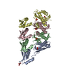
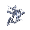
 Citation Citation | Journal: Science / Year: 2011 Title: Three-dimensional model of Salmonella's needle complex at subnanometer resolution. Authors: Oliver Schraidt / Thomas C Marlovits /  Abstract: Type III secretion systems (T3SSs) are essential virulence factors used by many Gram-negative bacteria to inject proteins that make eukaryotic host cells accessible to invasion. The T3SS core ...Type III secretion systems (T3SSs) are essential virulence factors used by many Gram-negative bacteria to inject proteins that make eukaryotic host cells accessible to invasion. The T3SS core structure, the needle complex (NC), is a ~3.5 megadalton-sized, oligomeric, membrane-embedded complex. Analyzing cryo-electron microscopy images of top views of NCs or NC substructures from Salmonella typhimurium revealed a 24-fold symmetry for the inner rings and a 15-fold symmetry for the outer rings, giving an overall C3 symmetry. Local refinement and averaging showed the organization of the central core and allowed us to reconstruct a subnanometer composite structure of the NC, which together with confident docking of atomic structures reveal insights into its overall organization and structural requirements during assembly. | |||||||||
| History |
|
- Structure visualization
| Movie |
Movie viewer |
|---|---|
| Structure viewer | EM map: SurfViewMolmilJmol/JSmol |
| Supplemental images |

- Downloads & links
-EMDB archive
| Map data | emd_1874.map.gz | 25.2 MB | EMDB map data format | |
|---|---|---|---|---|
| Header (meta data) | emd-1874-v30.xmlemd-1874.xml | 9.8 KB 9.8 KB | Display Display | EMDB header |
| Images | 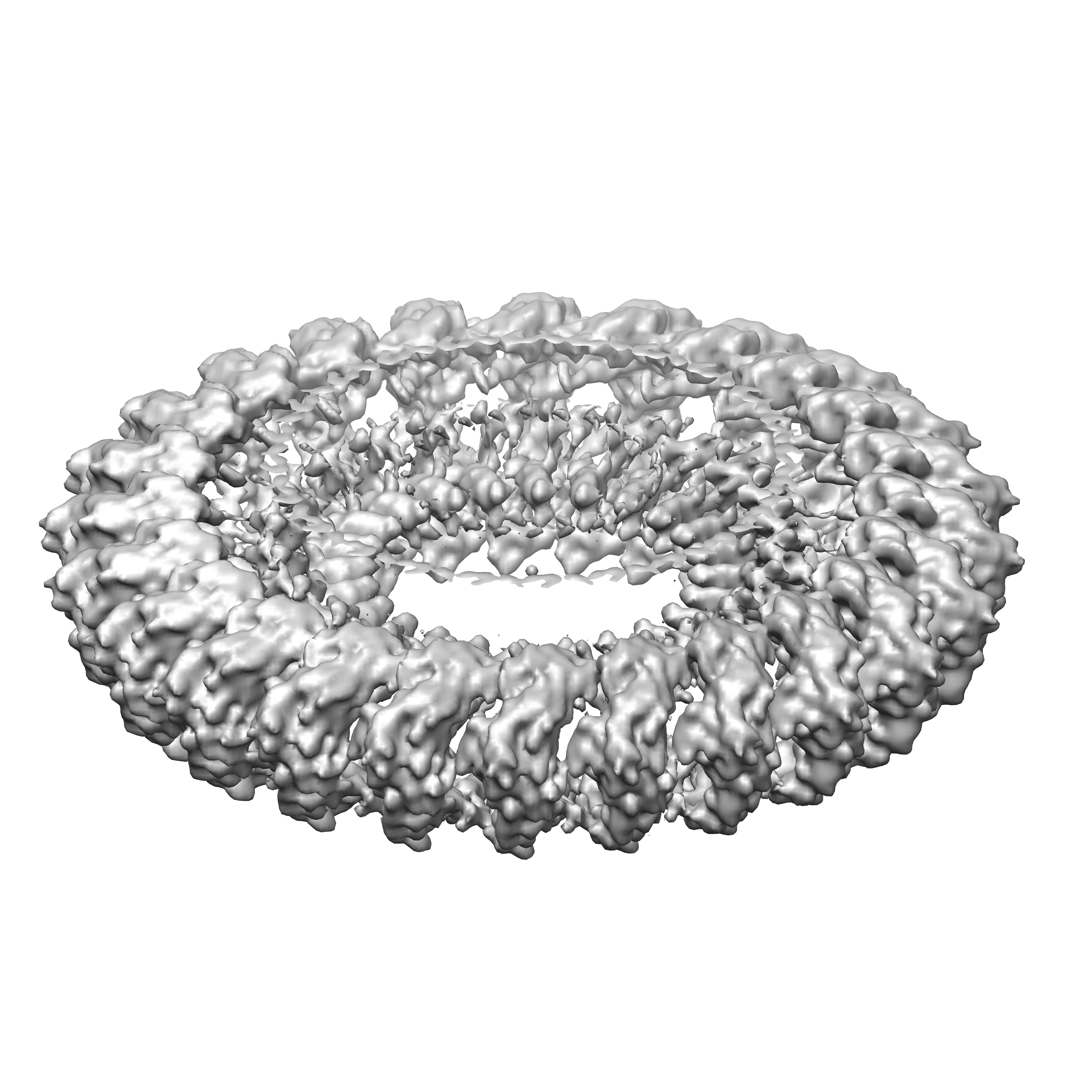 1874.jpg 1874.jpg | 1.1 MB | ||
| Archive directory |  http://ftp.pdbj.org/pub/emdb/structures/EMD-1874ftp://ftp.pdbj.org/pub/emdb/structures/EMD-1874 http://ftp.pdbj.org/pub/emdb/structures/EMD-1874ftp://ftp.pdbj.org/pub/emdb/structures/EMD-1874 | HTTPS FTP |
-Validation report
| Summary document | emd_1874_validation.pdf.gz | 244 KB | Display | EMDB validaton report |
|---|---|---|---|---|
| Full document | emd_1874_full_validation.pdf.gz | 243.1 KB | Display | |
| Data in XML | emd_1874_validation.xml.gz | 6.5 KB | Display | |
| Arichive directory | https://ftp.pdbj.org/pub/emdb/validation_reports/EMD-1874ftp://ftp.pdbj.org/pub/emdb/validation_reports/EMD-1874 | HTTPS FTP |
-Related structure data
| Related structure data | 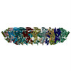 2y9jMC 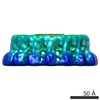 1871C 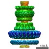 1875C  2y9kC C: citing same article ( M: atomic model generated by this map |
|---|---|
| Similar structure data |

-Links
| EMDB pages | EMDB (EBI/PDBe) / EMDataResource |
|---|---|
| Related items in Molecule of the Month |


-Map
| File | Download / File: emd_1874.map.gz / Format: CCP4 / Size: 100.6 MB / Type: IMAGE STORED AS FLOATING POINT NUMBER (4 BYTES) | ||||||||||||||||||||||||||||||||||||||||||||||||||||||||||||||||||||
|---|---|---|---|---|---|---|---|---|---|---|---|---|---|---|---|---|---|---|---|---|---|---|---|---|---|---|---|---|---|---|---|---|---|---|---|---|---|---|---|---|---|---|---|---|---|---|---|---|---|---|---|---|---|---|---|---|---|---|---|---|---|---|---|---|---|---|---|---|---|
| Annotation | This in an image of a surface rendered, extracted volume of the IR1 from the needle complex from Salmonella typhimurium | ||||||||||||||||||||||||||||||||||||||||||||||||||||||||||||||||||||
| Voxel size | X=Y=Z: 1.33 Å | ||||||||||||||||||||||||||||||||||||||||||||||||||||||||||||||||||||
| Density |
| ||||||||||||||||||||||||||||||||||||||||||||||||||||||||||||||||||||
| Symmetry | Space group: 1 | ||||||||||||||||||||||||||||||||||||||||||||||||||||||||||||||||||||
| Details | EMDB XML:
CCP4 map header:
| ||||||||||||||||||||||||||||||||||||||||||||||||||||||||||||||||||||
-Supplemental data
- Sample components
-Entire : Needle complex of the type III secretion system from Salmonella t...
| Entire | Name: Needle complex of the type III secretion system from Salmonella typhimurium - periplasmic domain of the inner membrane associated ring IR1 |
|---|---|
| Components |
|
-Supramolecule #1000: Needle complex of the type III secretion system from Salmonella t...
| Supramolecule | Name: Needle complex of the type III secretion system from Salmonella typhimurium - periplasmic domain of the inner membrane associated ring IR1 type: sample / ID: 1000 / Oligomeric state: 1 / Number unique components: 2 |
|---|
-Macromolecule #1: Protein PrgH
| Macromolecule | Name: Protein PrgH / type: protein_or_peptide / ID: 1 / Name.synonym: Protein PrgH / Oligomeric state: 24mer / Recombinant expression: No |
|---|---|
| Source (natural) | Organism: Salmonella enterica subsp. enterica serovar Typhimurium (bacteria) Location in cell: Membrane |
-Macromolecule #2: Protein PrgK
| Macromolecule | Name: Protein PrgK / type: protein_or_peptide / ID: 2 / Name.synonym: Protein PrgK / Oligomeric state: 24mer / Recombinant expression: No |
|---|---|
| Source (natural) | Organism: Salmonella enterica subsp. enterica serovar Typhimurium (bacteria) Location in cell: Membrane |
-Experimental details
-Structure determination
| Method | cryo EM |
|---|---|
 Processing Processing | single particle reconstruction |
| Aggregation state | particle |
-Sample preparation
| Buffer | pH: 7.5 / Details: 10mM Tris-HCl, 0.5M NaCl, 0.1% LDAO |
|---|---|
| Vitrification | Cryogen name: ETHANE / Instrument: OTHER |
- Electron microscopy
| Microscope | FEI POLARA 300 |
|---|---|
| Image recording | Digitization - Sampling interval: 15 µm / Bits/pixel: 8 |
| Electron beam | Acceleration voltage: 300 kV / Electron source:  FIELD EMISSION GUN FIELD EMISSION GUN |
| Electron optics | Illumination mode: FLOOD BEAM / Imaging mode: BRIGHT FIELD / Cs: 2 mm |
| Sample stage | Specimen holder: Eucentric / Specimen holder model: GATAN LIQUID NITROGEN |
| Experimental equipment |  Model: Tecnai Polara / Image courtesy: FEI Company |
-Image processing
| CTF correction | Details: Micrograph |
|---|---|
| Final reconstruction | Applied symmetry - Point group: C24 (24 fold cyclic) / Algorithm: OTHER / Resolution.type: BY AUTHOR / Resolution: 6.4 Å / Resolution method: FSC 0.5 CUT-OFF / Software - Name: IMAGIC |
-Atomic model buiding 1
| Initial model | PDB ID: |
|---|---|
| Software | Name: Chimera |
| Details | Protocol: Rigid Body. Based on the homolog EscJ (PDB entry above) a model was built for PrgK (Swissmodel) which was subsequently used for the fitting experiment (rigid body, Chimera), each chain was fitted separately |
| Refinement | Space: REAL / Protocol: RIGID BODY FIT / Target criteria: Correlation |
| Output model | PDB-2y9j: |