ribonuclease P activity / tRNA 5'-leader removal / box C/D methylation guide snoRNP complex / maturation of LSU-rRNA / maturation of LSU-rRNA from tricistronic rRNA transcript (SSU-rRNA, 5.8S rRNA, LSU-rRNA) / ribosomal large subunit biogenesis / maturation of SSU-rRNA / mRNA splicing, via spliceosome / 5S rRNA binding / large ribosomal subunit rRNA binding ...ribonuclease P activity / tRNA 5'-leader removal / box C/D methylation guide snoRNP complex / maturation of LSU-rRNA / maturation of LSU-rRNA from tricistronic rRNA transcript (SSU-rRNA, 5.8S rRNA, LSU-rRNA) / ribosomal large subunit biogenesis / maturation of SSU-rRNA / mRNA splicing, via spliceosome / 5S rRNA binding / large ribosomal subunit rRNA binding / cytosolic large ribosomal subunit / cytoplasmic translation / tRNA binding / rRNA binding / negative regulation of translation / ribosome / structural constituent of ribosome / translation / ribonucleoprotein complex / DNA repair / mRNA binding / nucleotide binding / DNA binding / zinc ion binding Similarity search - Function
Ribosomal protein L10, N-terminal RNA-binding domain / Ribosomal protein L11, N-terminal domain / Ribosomal protein L11/L12, N-terminal domain / Single Heli x bin / Non-ribosomal Peptide Synthetase Peptidyl Carrier Protein; Chain A - #60 / Ribosomal protein L30/L7, central domain / Ribosomal protein L39 / 50s Ribosomal Protein L19e, Chain O, domain 1 - #10 / N-terminal domain of TfIIb - #30 / Ribosomal protein L21 ...Ribosomal protein L10, N-terminal RNA-binding domain / Ribosomal protein L11, N-terminal domain / Ribosomal protein L11/L12, N-terminal domain / Single Heli x bin / Non-ribosomal Peptide Synthetase Peptidyl Carrier Protein; Chain A - #60 / Ribosomal protein L30/L7, central domain / Ribosomal protein L39 / 50s Ribosomal Protein L19e, Chain O, domain 1 - #10 / N-terminal domain of TfIIb - #30 / Ribosomal protein L21 / Ribosomal Protein L31e; Chain: W; / Ribosomal protein L31 / Nuclear Transport Factor 2; Chain: A, - #80 / Molecule: Ribosomal Protein L15; Chain: K; domain 1 / Molecule: Ribosomal Protein L15; Chain: K; domain 1 - #10 / 3-methyladenine DNA Glycosylase II; Chain A, domain 3 / Ribosomal protein L24 / Ribosomal protein L3; domain 3 / Ribosomal protein L3, domain 3 / Ribosomal protein L15e / Ribosomal protein L15 / Ribosomal Protein L15; Chain: K; domain 2 - #10 / Ribosomal Protein L3; Chain: B; domain 2, / Ribosomal Protein L3; Chain: B; domain 2, - #10 / Ribosomal protein L19e, domain 2 / Ribosomal protein L19e, domain 3 / 50s Ribosomal Protein L19e, Chain O, domain 1 / Ribosomal Protein L4; Chain: A; / Ribosomal protein L4/L1 / Helix Hairpins - #310 / 50S ribosomal protein L10, archaea / Ribosomal Protein L13p; Chain: A; / Ribosomal protein L13 / Atp Synthase Epsilon Chain; Chain: I; / Ribosomal Protein L24e; Chain: T; / Ribosomal Protein L15; Chain: K; domain 2 / Ribosomal Protein L15; Chain: K; domain 2 / Ribosomal protein L19e, archaeal / Ribosomal protein L2; domain 3 / Ribosomal protein L2, domain 3 / Ribosomal protein L11/L12, C-terminal domain / Ribosomal protein L15e, archaeal / Ribosomal protein L30, archaeal / Ribosomal protein L6P, archaea / Ribosomal protein L14P, archaeal / Ribosomal protein L21e, archaeal / Ribosomal protein L18e, archaea / Ribosomal protein L10e, archaea / Ribosomal protein L32e, archaeal / Ribosomal protein L3, archaeal / Ribosomal protein L4, archaea / Ribosomal protein L5, archaeal / Ribosomal protein L6 / 50s Ribosomal Protein L5; Chain: A, / Ribosomal protein L5 / Ribosomal protein L30/L7 / Nucleotidyltransferase; domain 5 - #100 / Ribosomal protein L16/L10 / Ribosomal protein L22/L17 / Ribosomal Protein L14 / Ribosomal protein L14/L23 / Outer Surface Protein A; domain 3 / Ribosomal protein L7Ae, archaea / Ribosomal Protein L22; Chain A / Ribosomal Protein L30; Chain: A, / Ribosomal protein L24e / SH3 type barrels. - #30 / Ribosomal protein L30/S12 / Aldehyde Oxidoreductase; domain 3 / DNA repair Rad51/transcription factor NusA, alpha-helical / : / Non-ribosomal Peptide Synthetase Peptidyl Carrier Protein; Chain A / N-terminal domain of TfIIb / Helix-hairpin-helix domain / Ribosomal protein L2, archaeal-type / Ribosomal L15/L27a, N-terminal / Translation factors / : / Ribosomal protein L23 / 50S ribosomal protein L10, insertion domain superfamily / 60S ribosomal protein L10P, insertion domain / Insertion domain in 60S ribosomal protein L10P / metallochaperone-like domain / TRASH domain / RRM (RNA recognition motif) domain / 60s Ribosomal Protein L30; Chain: A; / : / Ribosomal protein L44e signature. / Helix-hairpin-helix DNA-binding motif, class 1 / Helix-hairpin-helix DNA-binding motif class 1 / Ribosomal protein L10e, conserved site / Ribosomal protein L10e signature. / Ribosomal protein L10e / Ribosomal protein L24e, conserved site / Ribosomal protein L24e signature. / Ribosomal protein L44e / Ribosomal protein L19/L19e conserved site / Ribosomal protein L44 / Ribosomal protein L19e signature. / Ribosomal protein L39e, conserved site Similarity search - Domain/homology
: / : / : / RNA / RNA (> 10) / RNA (> 100) / RNA (> 1000) / Large ribosomal subunit protein uL22 / Large ribosomal subunit protein uL29 / Large ribosomal subunit protein uL24 ...: / : / : / RNA / RNA (> 10) / RNA (> 100) / RNA (> 1000) / Large ribosomal subunit protein uL22 / Large ribosomal subunit protein uL29 / Large ribosomal subunit protein uL24 / Large ribosomal subunit protein uL23 / Large ribosomal subunit protein eL18 / Large ribosomal subunit protein eL21 / Large ribosomal subunit protein uL4 / Large ribosomal subunit protein eL32 / Large ribosomal subunit protein uL15 / Large ribosomal subunit protein eL8 / Large ribosomal subunit protein eL24 / Large ribosomal subunit protein eL19 / Large ribosomal subunit protein uL30 / Large ribosomal subunit protein uL11 / Large ribosomal subunit protein uL18 / Large ribosomal subunit protein uL5 / Large ribosomal subunit protein uL6 / Large ribosomal subunit protein uL10 / Large ribosomal subunit protein eL31 / Large ribosomal subunit protein uL2 / Large ribosomal subunit protein uL3 / Large ribosomal subunit protein uL14 / Large ribosomal subunit protein eL39 / Large ribosomal subunit protein uL13 / Large ribosomal subunit protein eL37 / Large ribosomal subunit protein eL42 / Large ribosomal subunit protein uL16 / Large ribosomal subunit protein eL15 / Large ribosomal subunit protein eL43 Similarity search - Component
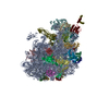
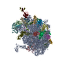
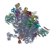
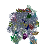
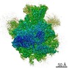
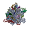
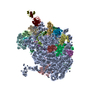
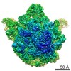
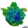
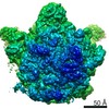
Biological species
Haloarcula marismortui (Halophile)
Method
X-RAY DIFFRACTION / SYNCHROTRON / CNS / Resolution: 3 Å
Mass: 112.411 Da / Num. of mol.: 5 / Source method: obtained synthetically / Formula: Cd
-
Details
Sequence details
RESIDUE A 1207 AND RESIDUE C 1208 CHAIN 0 ARE NOT LINKED. DISTANCE OF O3'-P BOND IS 2.51A. DISTANCE ...RESIDUE A 1207 AND RESIDUE C 1208 CHAIN 0 ARE NOT LINKED. DISTANCE OF O3'-P BOND IS 2.51A. DISTANCE OF C-N BOND BETWEEN RESIDUE G LEU 53 AND RESIDUE G HIS 54 IS 1.63A. DISTANCE OF C-N BOND BETWEEN RESIDUE G HIS 54 AND RESIDUE G GLY 55 IS 0.99A.
-
Experimental details
-
Experiment
Experiment
Method: X-RAY DIFFRACTION / Number of used crystals: 1
-
Sample preparation
Crystal
Density Matthews: 3.18 Å3/Da / Density % sol: 61.31 %
In the structure databanks used in Yorodumi, some data are registered as the other names, "COVID-19 virus" and "2019-nCoV". Here are the details of the virus and the list of structure data.
Jan 31, 2019. EMDB accession codes are about to change! (news from PDBe EMDB page)
EMDB accession codes are about to change! (news from PDBe EMDB page)
The allocation of 4 digits for EMDB accession codes will soon come to an end. Whilst these codes will remain in use, new EMDB accession codes will include an additional digit and will expand incrementally as the available range of codes is exhausted. The current 4-digit format prefixed with “EMD-” (i.e. EMD-XXXX) will advance to a 5-digit format (i.e. EMD-XXXXX), and so on. It is currently estimated that the 4-digit codes will be depleted around Spring 2019, at which point the 5-digit format will come into force.
The EM Navigator/Yorodumi systems omit the EMD- prefix.
Related info.:Q: What is EMD? / ID/Accession-code notation in Yorodumi/EM Navigator
Yorodumi is a browser for structure data from EMDB, PDB, SASBDB, etc.
This page is also the successor to EM Navigator detail page, and also detail information page/front-end page for Omokage search.
The word "yorodu" (or yorozu) is an old Japanese word meaning "ten thousand". "mi" (miru) is to see.
Related info.:EMDB / PDB / SASBDB / Comparison of 3 databanks / Yorodumi Search / Aug 31, 2016. New EM Navigator & Yorodumi / Yorodumi Papers / Jmol/JSmol / Function and homology information / Changes in new EM Navigator and Yorodumi
 Movie
Movie Controller

 Yorodumi
 Open data
 Basic information
 Components
 Keywords
 Function and homology information
 Haloarcula marismortui (Halophile)
 X-RAY DIFFRACTION /
  Authors
 Citation
 Structure visualization
 Downloads & links
 Other downloads
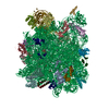
 PDBj
PDBj


 Assembly


 Mass: 24.305 Da / Num. of mol.: 116 / Source method: obtained synthetically / Formula: Mg
 Mass: 39.098 Da / Num. of mol.: 2 / Source method: obtained synthetically / Formula: K
 Mass: 22.990 Da / Num. of mol.: 86 / Source method: obtained synthetically / Formula: Na
 Mass: 35.453 Da / Num. of mol.: 22 / Source method: obtained synthetically / Formula: Cl
 Mass: 112.411 Da / Num. of mol.: 5 / Source method: obtained synthetically / Formula: Cd
 Sample preparation
 / Beamline: 24-ID-C / Wavelength: 0.979 Å
 Processing