- EMDB-3133: Cryo-EM structures of the 50S ribosome subunit bound with HflX -
+
Open data
ID or keywords:
Loading...
-
Basic information
Entry
Database: EMDB / ID: EMD-3133
Title
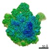
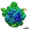
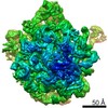
Cryo-EM structures of the 50S ribosome subunit bound with HflX
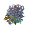
Map data
Cryo-EM structures of the 50S ribosome subunit bound with HflX
Sample
Sample: 50S-HflX complex
Complex: prokaryotic 50S ribosome subunit
Protein or peptide: HflX
Keywords
Ribosome rescue
Function / homology
Function and homology information
ribosome disassembly / guanosine tetraphosphate binding / stringent response / ribosomal large subunit binding / transcriptional attenuation / endoribonuclease inhibitor activity / RNA-binding transcription regulator activity / positive regulation of ribosome biogenesis / negative regulation of cytoplasmic translation / translational termination ...ribosome disassembly / guanosine tetraphosphate binding / stringent response / ribosomal large subunit binding / transcriptional attenuation / endoribonuclease inhibitor activity / RNA-binding transcription regulator activity / positive regulation of ribosome biogenesis / negative regulation of cytoplasmic translation / translational termination / DnaA-L2 complex / translation repressor activity / negative regulation of translational initiation / negative regulation of DNA-templated DNA replication initiation / rescue of stalled ribosome / mRNA regulatory element binding translation repressor activity / ribosome assembly / assembly of large subunit precursor of preribosome / cytosolic ribosome assembly / ribosomal large subunit assembly / response to reactive oxygen species / regulation of cell growth / DNA-templated transcription termination / response to radiation / mRNA 5'-UTR binding / large ribosomal subunit / ribosome binding / transferase activity / 5S rRNA binding / large ribosomal subunit rRNA binding / response to heat / cytosolic large ribosomal subunit / cytoplasmic translation / tRNA binding / rRNA binding / negative regulation of translation / ribosome / structural constituent of ribosome / translation / response to antibiotic / negative regulation of DNA-templated transcription / GTPase activity / mRNA binding / GTP binding / ATP hydrolysis activity / DNA binding / RNA binding / zinc ion binding / ATP binding / metal ion binding / cytoplasm / cytosol Similarity search - Function
HflX, C-terminal domain / HflX C-terminal domain / GTPase HflX / GTPase HflX, N-terminal / HflX-type guanine nucleotide-binding (G) domain / GTP-binding protein, middle domain / GTPase HflX, N-terminal domain superfamily / GTP-binding GTPase N-terminal / GTP-binding GTPase Middle Region / HflX-type guanine nucleotide-binding (G) domain profile. ...HflX, C-terminal domain / HflX C-terminal domain / GTPase HflX / GTPase HflX, N-terminal / HflX-type guanine nucleotide-binding (G) domain / GTP-binding protein, middle domain / GTPase HflX, N-terminal domain superfamily / GTP-binding GTPase N-terminal / GTP-binding GTPase Middle Region / HflX-type guanine nucleotide-binding (G) domain profile. / 50S ribosome-binding GTPase / Ribosomal protein L1, bacterial-type / GTP binding domain / Ribosomal protein L10, eubacterial, conserved site / Ribosomal protein L10 signature. / Ribosomal protein L10 / EF-G domain III/V-like / : / Ribosomal protein L1, conserved site / Ribosomal protein L1 signature. / Ribosomal protein L1 / Ribosomal protein L1, 3-layer alpha/beta-sandwich / Ribosomal protein L25, short-form / Ribosomal protein L1-like / Ribosomal protein L1/ribosomal biogenesis protein / Ribosomal protein L1p/L10e family / Ribosomal protein L11, bacterial-type / : / Ribosomal protein L21, conserved site / Ribosomal protein L21 signature. / Ribosomal protein L11, conserved site / Ribosomal protein L11 signature. / Ribosomal protein L10-like domain superfamily / Ribosomal protein L16 signature 1. / : / Ribosomal protein L10 / Ribosomal protein L10P / Ribosomal protein L6, conserved site / Ribosomal protein L6 signature 1. / Ribosomal protein L16, conserved site / Ribosomal protein L16 signature 2. / Ribosomal protein L9 signature. / Ribosomal protein L9, bacteria/chloroplast / Ribosomal protein L9, C-terminal / Ribosomal protein L9, C-terminal domain / Ribosomal protein L9, C-terminal domain superfamily / Ribosomal protein L17 signature. / Ribosomal L25p family / Ribosomal protein L25 / Ribosomal protein L11, N-terminal / Ribosomal protein L11, N-terminal domain / Ribosomal protein L11/L12 / Ribosomal protein L11, C-terminal / Ribosomal protein L11, C-terminal domain superfamily / Ribosomal protein L11/L12, N-terminal domain superfamily / Ribosomal protein L11, RNA binding domain / Ribosomal protein L11/L12 / Ribosomal protein L36 signature. / Ribosomal protein L28/L24 superfamily / Ribosomal protein L25/Gln-tRNA synthetase, N-terminal / Ribosomal protein L32p, bacterial type / Ribosomal protein L25/Gln-tRNA synthetase, anti-codon-binding domain superfamily / : / Ribosomal protein L9, N-terminal domain superfamily / Ribosomal protein L9 / Ribosomal protein L9, N-terminal / Ribosomal protein L9, N-terminal domain / : / Ribosomal protein L35, conserved site / Ribosomal protein L35 signature. / Ribosomal protein L28 / Ribosomal protein L33, conserved site / Ribosomal protein L33 signature. / Ribosomal protein L35, non-mitochondrial / Ribosomal protein L5, bacterial-type / Ribosomal protein L18, bacterial-type / Ribosomal protein L6, bacterial-type / Ribosomal protein L19, conserved site / Ribosomal protein L19 signature. / Ribosomal protein L9/RNase H1, N-terminal / Ribosomal protein L36 / Ribosomal protein L36 superfamily / Ribosomal protein L36 / Ribosomal protein L20 signature. / Ribosomal protein L27, conserved site / Ribosomal protein L27 signature. / Ribosomal protein L14P, bacterial-type / Ribosomal protein L34, conserved site / Ribosomal protein L34 signature. / Ribosomal protein L22, bacterial/chloroplast-type / Ribosomal protein L2, bacterial/organellar-type / Ribosomal protein L35 / Ribosomal protein L35 superfamily / Ribosomal protein L35 / Ribosomal L28 family / Ribosomal protein L33 / Ribosomal protein L33 / Ribosomal protein L28/L24 / Ribosomal protein L18 / Ribosomal L18 of archaea, bacteria, mitoch. and chloroplast Similarity search - Domain/homology
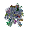
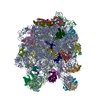
Large ribosomal subunit protein uL15 / Large ribosomal subunit protein uL10 / Large ribosomal subunit protein uL11 / Large ribosomal subunit protein bL19 / Large ribosomal subunit protein uL1 / Large ribosomal subunit protein bL20 / Large ribosomal subunit protein bL27 / Large ribosomal subunit protein bL28 / Large ribosomal subunit protein uL29 / Large ribosomal subunit protein bL32 ...Large ribosomal subunit protein uL15 / Large ribosomal subunit protein uL10 / Large ribosomal subunit protein uL11 / Large ribosomal subunit protein bL19 / Large ribosomal subunit protein uL1 / Large ribosomal subunit protein bL20 / Large ribosomal subunit protein bL27 / Large ribosomal subunit protein bL28 / Large ribosomal subunit protein uL29 / Large ribosomal subunit protein bL32 / Large ribosomal subunit protein bL33 / Large ribosomal subunit protein bL34 / Large ribosomal subunit protein bL35 / Large ribosomal subunit protein bL36A / Large ribosomal subunit protein bL9 / Large ribosomal subunit protein uL13 / Large ribosomal subunit protein uL14 / Large ribosomal subunit protein uL16 / Large ribosomal subunit protein uL23 / Large ribosomal subunit protein bL17 / Large ribosomal subunit protein bL21 / Large ribosomal subunit protein uL30 / Large ribosomal subunit protein uL6 / Large ribosomal subunit protein uL18 / GTPase HflX / Large ribosomal subunit protein uL2 / Large ribosomal subunit protein uL3 / Large ribosomal subunit protein uL24 / Large ribosomal subunit protein uL4 / Large ribosomal subunit protein uL22 / Large ribosomal subunit protein uL5 / Large ribosomal subunit protein bL25 Similarity search - Component
Biological species
Escherichia coli K-12 (bacteria) / Escherichia coli (E. coli)
Method
single particle reconstruction / cryo EM / Resolution: 4.5 Å
Journal: Nat Struct Mol Biol / Year: 2015 Title: HflX is a ribosome-splitting factor rescuing stalled ribosomes under stress conditions. Authors: Yanqing Zhang / Chandra Sekhar Mandava / Wei Cao / Xiaojing Li / Dejiu Zhang / Ningning Li / Yixiao Zhang / Xiaoxiao Zhang / Yan Qin / Kaixia Mi / Jianlin Lei / Suparna Sanyal / Ning Gao / Abstract: Adverse cellular conditions often lead to nonproductive translational stalling and arrest of ribosomes on mRNAs. Here, we used fast kinetics and cryo-EM to characterize Escherichia coli HflX, a ...Adverse cellular conditions often lead to nonproductive translational stalling and arrest of ribosomes on mRNAs. Here, we used fast kinetics and cryo-EM to characterize Escherichia coli HflX, a GTPase with unknown function. Our data reveal that HflX is a heat shock-induced ribosome-splitting factor capable of dissociating vacant as well as mRNA-associated ribosomes with deacylated tRNA in the peptidyl site. Structural data demonstrate that the N-terminal effector domain of HflX binds to the peptidyl transferase center in a strikingly similar manner as that of the class I release factors and induces dramatic conformational changes in central intersubunit bridges, thus promoting subunit dissociation. Accordingly, loss of HflX results in an increase in stalled ribosomes upon heat shock. These results suggest a primary role of HflX in rescuing translationally arrested ribosomes under stress conditions.
History
Deposition
Aug 23, 2015
-
Header (metadata) release
Sep 16, 2015
-
Map release
Oct 14, 2015
-
Update
Dec 2, 2015
-
Current status
Dec 2, 2015
Processing site: PDBe / Status: Released
-
Structure visualization
Movie
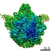
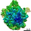
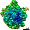
Surface view with section colored by density value
Organism: Escherichia coli K-12 (bacteria) / Location in cell: Cytoplasm
Molecular weight
Experimental: 1.5 MDa / Theoretical: 1.5 MDa
-
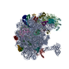
Macromolecule #1: HflX
Macromolecule
Name: HflX / type: protein_or_peptide / ID: 1 Details: GMPPNP was bound with HflX in the 50S-HflX complex. Number of copies: 1 / Oligomeric state: monomer / Recombinant expression: Yes
Source (natural)
Organism: Escherichia coli (E. coli) / Location in cell: Cytoplasm
Atomic model of E. coli HflX was modeled from the crystal structure of Sulfolobus solfataricus HflX (PDB id: 3KXI).The homology modeling was performed with MODELLER. The model of the CTD of E. coli HflX was independently modeled by I-TASSER (template PDB code: 2WBM, residues 164-232). The switch I region disordered in the crystal structure of S. solfataricus HflX was modeled using the crystal structure of S. thermophilus NFeoB (PDB id: 3B1X) as a template. GMPPNP was derived from a previous model (PDB id: 3B1X) and docked into the atomic model of the E. coli HflX.
Refinement
Space: REAL / Protocol: FLEXIBLE FIT
Output model
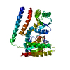
PDB-5ady: Cryo-EM structures of the 50S ribosome subunit bound with HflX
+
About Yorodumi
-
News
-
Feb 9, 2022. New format data for meta-information of EMDB entries
New format data for meta-information of EMDB entries
Version 3 of the EMDB header file is now the official format.
The previous official version 1.9 will be removed from the archive.
In the structure databanks used in Yorodumi, some data are registered as the other names, "COVID-19 virus" and "2019-nCoV". Here are the details of the virus and the list of structure data.
Jan 31, 2019. EMDB accession codes are about to change! (news from PDBe EMDB page)
EMDB accession codes are about to change! (news from PDBe EMDB page)
The allocation of 4 digits for EMDB accession codes will soon come to an end. Whilst these codes will remain in use, new EMDB accession codes will include an additional digit and will expand incrementally as the available range of codes is exhausted. The current 4-digit format prefixed with “EMD-” (i.e. EMD-XXXX) will advance to a 5-digit format (i.e. EMD-XXXXX), and so on. It is currently estimated that the 4-digit codes will be depleted around Spring 2019, at which point the 5-digit format will come into force.
The EM Navigator/Yorodumi systems omit the EMD- prefix.
Related info.:Q: What is EMD? / ID/Accession-code notation in Yorodumi/EM Navigator
Yorodumi is a browser for structure data from EMDB, PDB, SASBDB, etc.
This page is also the successor to EM Navigator detail page, and also detail information page/front-end page for Omokage search.
The word "yorodu" (or yorozu) is an old Japanese word meaning "ten thousand". "mi" (miru) is to see.
Related info.:EMDB / PDB / SASBDB / Comparison of 3 databanks / Yorodumi Search / Aug 31, 2016. New EM Navigator & Yorodumi / Yorodumi Papers / Jmol/JSmol / Function and homology information / Changes in new EM Navigator and Yorodumi
 Movie
Movie Controller


 Open data
 Basic information
 Map data
 Sample
 Keywords
 Function and homology information
 Authors
 Citation

 Structure visualization
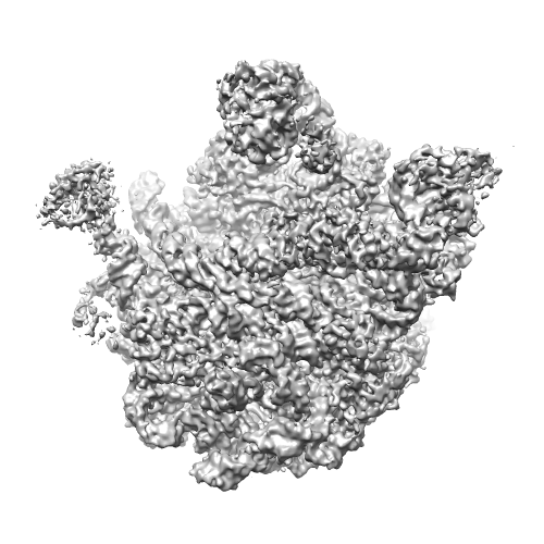
 Downloads & links
 emd_3133.png
 http://ftp.pdbj.org/pub/emdb/structures/EMD-3133
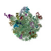


 Sample components
 Processing
 Electron microscopy
 FIELD EMISSION GUN