ムービー
ムービー コントローラー


+ データを開く

- 基本情報
基本情報
| 登録情報 | データベース: EMDB / ID: EMD-1255 | |||||||||
|---|---|---|---|---|---|---|---|---|---|---|
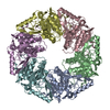
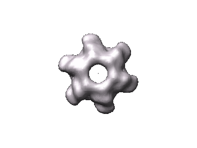
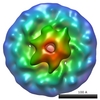
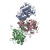
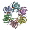
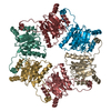
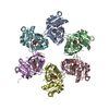
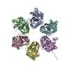
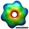
| タイトル | Structure of a hexameric RNA packaging motor in a viral polymerase complex. | |||||||||
 マップデータ マップデータ | Bacteriophage phi8 hexameric packaging motor P4 reconstructed within the viral cores (icosahedral contribution of the core subtracted in the images), applying C6 symmetry | |||||||||
 試料 試料 |
| |||||||||
| 生物種 |  Pseudomonas phage phi8 (ファージ) Pseudomonas phage phi8 (ファージ) | |||||||||
| 手法 |  単粒子再構成法 / クライオ電子顕微鏡法 / 解像度: 15.0 Å 単粒子再構成法 / クライオ電子顕微鏡法 / 解像度: 15.0 Å | |||||||||
 データ登録者 データ登録者 | Huiskonen JT / Jaalinoja HT / Briggs JAG | |||||||||
 引用 引用 | ジャーナル: J Struct Biol / 年: 2007 タイトル: Structure of a hexameric RNA packaging motor in a viral polymerase complex. 著者: Juha T Huiskonen / Harri T Jäälinoja / John A G Briggs / Stephen D Fuller / Sarah J Butcher /  要旨: Packaging of the Cystovirus varphi8 genome into the polymerase complex is catalysed by the hexameric P4 packaging motor. The motor is located at the fivefold vertices of the icosahedrally symmetric ...Packaging of the Cystovirus varphi8 genome into the polymerase complex is catalysed by the hexameric P4 packaging motor. The motor is located at the fivefold vertices of the icosahedrally symmetric polymerase complex, and the symmetry mismatch between them may be critical for function. We have developed a novel image-processing approach for the analysis of symmetry-mismatched structures and applied it to cryo-electron microscopy images of P4 bound to the polymerase complex. This approach allowed us to solve the three-dimensional structure of the P4 in situ to 15-A resolution. The C-terminal face of P4 was observed to interact with the polymerase complex, supporting the current view on RNA translocation. We suggest that the symmetry mismatch between the two components may facilitate the ring opening required for RNA loading prior to its translocation. | |||||||||
| 履歴 |
|
- 構造の表示
構造の表示
| ムービー |
 ムービービューア ムービービューア |
|---|---|
| 構造ビューア | EMマップ: SurfViewMolmilJmol/JSmol |
| 添付画像 |
- ダウンロードとリンク
-EMDBアーカイブ
| マップデータ | emd_1255.map.gz | 3.3 MB | EMDBマップデータ形式 | |
|---|---|---|---|---|
| ヘッダ (付随情報) | emd-1255-v30.xmlemd-1255.xml | 8.9 KB 8.9 KB | 表示 表示 | EMDBヘッダ |
| 画像 |  1255.gif 1255.gif | 10.9 KB | ||
| アーカイブディレクトリ |  http://ftp.pdbj.org/pub/emdb/structures/EMD-1255ftp://ftp.pdbj.org/pub/emdb/structures/EMD-1255 http://ftp.pdbj.org/pub/emdb/structures/EMD-1255ftp://ftp.pdbj.org/pub/emdb/structures/EMD-1255 | HTTPS FTP |
-関連構造データ
-リンク
| EMDBのページ | EMDB (EBI/PDBe) / EMDataResource |
|---|
-マップ
| ファイル | ダウンロード / ファイル: emd_1255.map.gz / 形式: CCP4 / 大きさ: 3.7 MB / タイプ: IMAGE STORED AS FLOATING POINT NUMBER (4 BYTES) | ||||||||||||||||||||||||||||||||||||||||||||||||||||||||||||||||||||
|---|---|---|---|---|---|---|---|---|---|---|---|---|---|---|---|---|---|---|---|---|---|---|---|---|---|---|---|---|---|---|---|---|---|---|---|---|---|---|---|---|---|---|---|---|---|---|---|---|---|---|---|---|---|---|---|---|---|---|---|---|---|---|---|---|---|---|---|---|---|
| 注釈 | Bacteriophage phi8 hexameric packaging motor P4 reconstructed within the viral cores (icosahedral contribution of the core subtracted in the images), applying C6 symmetry | ||||||||||||||||||||||||||||||||||||||||||||||||||||||||||||||||||||
| ボクセルのサイズ | X=Y=Z: 2.8 Å | ||||||||||||||||||||||||||||||||||||||||||||||||||||||||||||||||||||
| 密度 |
| ||||||||||||||||||||||||||||||||||||||||||||||||||||||||||||||||||||
| 対称性 | 空間群: 1 | ||||||||||||||||||||||||||||||||||||||||||||||||||||||||||||||||||||
| 詳細 | EMDB XML:
CCP4マップ ヘッダ情報:
| ||||||||||||||||||||||||||||||||||||||||||||||||||||||||||||||||||||
-添付データ
- 試料の構成要素
-全体 : Bacteriophage phi8 core
| 全体 | 名称: Bacteriophage phi8 core |
|---|---|
| 要素 |
|
-超分子 #1000: Bacteriophage phi8 core
| 超分子 | 名称: Bacteriophage phi8 core / タイプ: sample / ID: 1000 / Number unique components: 1 |
|---|---|
| 分子量 | 理論値: 33 MDa |
-超分子 #1: Pseudomonas phage phi8
| 超分子 | 名称: Pseudomonas phage phi8 / タイプ: virus / ID: 1 / Name.synonym: Bacteriophage phi8 / NCBI-ID: 120086 / 生物種: Pseudomonas phage phi8 / ウイルスタイプ: VIRION / ウイルス・単離状態: SPECIES / ウイルス・エンベロープ: Yes / ウイルス・中空状態: No / Syn species name: Bacteriophage phi8 |
|---|---|
| 宿主 | 生物種:  Pseudomonas syringae pv. phaseolicola (バクテリア) Pseudomonas syringae pv. phaseolicola (バクテリア)別称: BACTERIA(EUBACTERIA) |
| 分子量 | 実験値: 33 MDa |
| ウイルス殻 | Shell ID: 1 / T番号(三角分割数): 1 |
-実験情報
-構造解析
| 手法 | クライオ電子顕微鏡法 |
|---|---|
 解析 解析 | 単粒子再構成法 |
| 試料の集合状態 | particle |
-試料調製
| 緩衝液 | pH: 7.5 詳細: 10 mM potassium phosphate pH 7.5, 1 mM MgCl2, 50 mM NaCl |
|---|---|
| グリッド | 詳細: 200 mesh holey carbon coated copper grid |
| 凍結 | 凍結剤: ETHANE |
- 電子顕微鏡法
| 顕微鏡 | FEI TECNAI F20 |
|---|---|
| 電子線 | 加速電圧: 200 kV / 電子線源: FIELD EMISSION GUN |
| 電子光学系 | 照射モード: FLOOD BEAM / 撮影モード: BRIGHT FIELDBright-field microscopy / Cs: 2.0 mm / 最大 デフォーカス(公称値): 3.3 µm / 最小 デフォーカス(公称値): 0.7 µm / 倍率(公称値): 50000 |
| 試料ステージ | 試料ホルダー: Side entry liquid nitrogen-cooled cryo specimen holder 試料ホルダーモデル: OTHER |
| 撮影 | カテゴリ: FILM / フィルム・検出器のモデル: KODAK SO-163 FILM / デジタル化 - スキャナー: ZEISS SCAI / デジタル化 - サンプリング間隔: 7 µm / 実像数: 66 / ビット/ピクセル: 12 |
| 実験機器 |  モデル: Tecnai F20 / 画像提供: FEI Company |
-画像解析
| CTF補正 | 詳細: Each particle, phases flipped |
|---|---|
| 最終 2次元分類 | クラス数: 352 |
| 最終 再構成 | 想定した対称性 - 点群: C6 (6回回転対称) / アルゴリズム: OTHER / 解像度のタイプ: BY AUTHOR / 解像度: 15.0 Å / 解像度の算出法: FSC 3 SIGMA CUT-OFF / ソフトウェア - 名称: Imagic5 |
-原子モデル構築 1
| 初期モデル | PDB ID: |
|---|---|
| ソフトウェア | 名称: colores |
| 詳細 | Protocol: rigid body |
| 精密化 | 空間: RECIPROCAL / プロトコル: RIGID BODY FIT / 当てはまり具合の基準: correlation coefficient |