 ムービー
ムービー コントローラー


+ データを開く

- 基本情報
基本情報
| 登録情報 | データベース: PDB / ID: 8ihj | ||||||
|---|---|---|---|---|---|---|---|
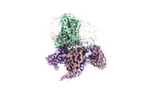
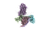
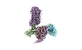
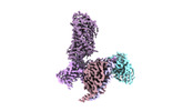
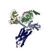
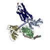
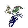
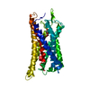
| タイトル | Cryo-EM structure of HCA3-Gi complex with acifran | ||||||
 要素 要素 |
| ||||||
 キーワード キーワード |  SIGNALING PROTEIN / GPCR (Gタンパク質共役受容体) SIGNALING PROTEIN / GPCR (Gタンパク質共役受容体) | ||||||
| 機能・相同性 |  機能・相同性情報 機能・相同性情報Activation of the phototransduction cascade / Sensory perception of sweet, bitter, and umami (glutamate) taste / Olfactory Signaling Pathway / G beta:gamma signalling through PLC beta / Presynaptic function of Kainate receptors / Prostacyclin signalling through prostacyclin receptor / G alpha (z) signalling events / Glucagon-type ligand receptors / G beta:gamma signalling through PI3Kgamma / G beta:gamma signalling through CDC42 ...Activation of the phototransduction cascade / Sensory perception of sweet, bitter, and umami (glutamate) taste / Olfactory Signaling Pathway / G beta:gamma signalling through PLC beta / Presynaptic function of Kainate receptors / Prostacyclin signalling through prostacyclin receptor / G alpha (z) signalling events / Glucagon-type ligand receptors / G beta:gamma signalling through PI3Kgamma / G beta:gamma signalling through CDC42 / Synthesis, secretion, and inactivation of Glucagon-like Peptide-1 (GLP-1) / G beta:gamma signalling through BTK / Adrenaline,noradrenaline inhibits insulin secretion / ADP signalling through P2Y purinoceptor 12 / Cooperation of PDCL (PhLP1) and TRiC/CCT in G-protein beta folding / Thromboxane signalling through TP receptor / Thrombin signalling through proteinase activated receptors (PARs) / Activation of G protein gated Potassium channels / Inhibition of voltage gated Ca2+ channels via Gbeta/gamma subunits / G-protein activation / nicotinic acid receptor activity / Hydroxycarboxylic acid-binding receptors / Ca2+ pathway / G alpha (s) signalling events / G alpha (q) signalling events / Extra-nuclear estrogen signaling / G alpha (12/13) signalling events / Vasopressin regulates renal water homeostasis via Aquaporins / G alpha (i) signalling events / GPER1 signaling / Glucagon-like Peptide-1 (GLP1) regulates insulin secretion / ADP signalling through P2Y purinoceptor 1 / phototransduction, visible light / alkylglycerophosphoethanolamine phosphodiesterase activity / photoreceptor outer segment membrane / spectrin binding / photoreceptor outer segment / Adenylate cyclase inhibitory pathway / positive regulation of protein localization to cell cortex / regulation of cAMP-mediated signaling / D2 dopamine receptor binding / G protein-coupled serotonin receptor binding / regulation of mitotic spindle organization / cellular response to forskolin / cardiac muscle cell apoptotic process / adenylate cyclase-inhibiting G protein-coupled receptor signaling pathway / photoreceptor inner segment / Regulation of insulin secretion / G protein-coupled receptor binding / G protein-coupled receptor activity / G-protein beta/gamma-subunit complex binding / adenylate cyclase-modulating G protein-coupled receptor signaling pathway / ADP signalling through P2Y purinoceptor 12 / response to peptide hormone / Adrenaline,noradrenaline inhibits insulin secretion / G alpha (z) signalling events / ADORA2B mediated anti-inflammatory cytokines production / sensory perception of taste / GPER1 signaling / G-protein beta-subunit binding / GDP binding / heterotrimeric G-protein complex / signaling receptor complex adaptor activity / retina development in camera-type eye / 細胞結合 / GTPase binding / 髄鞘 / cell body / phospholipase C-activating G protein-coupled receptor signaling pathway / 細胞皮質 / midbody / G alpha (i) signalling events / positive regulation of cytosolic calcium ion concentration / fibroblast proliferation / cellular response to hypoxia / G alpha (s) signalling events / cell population proliferation / Extra-nuclear estrogen signaling / electron transfer activity / ペリプラズム / 細胞周期 / iron ion binding / G protein-coupled receptor signaling pathway / lysosomal membrane / 細胞分裂 / GTPase activity / 中心体 / 樹状突起 / heme binding / protein-containing complex binding / GTP binding / 核小体 / magnesium ion binding / extracellular exosome / 核質 / 細胞膜 / 細胞質類似検索 - 分子機能 | ||||||
| 生物種 |  Escherichia coli (大腸菌) Escherichia coli (大腸菌) Homo sapiens (ヒト) Homo sapiens (ヒト)synthetic construct (人工物)  Mus musculus (ハツカネズミ) Mus musculus (ハツカネズミ) | ||||||
| 手法 | 電子顕微鏡法 / 単粒子再構成法 / クライオ電子顕微鏡法 / 解像度: 3.07 Å | ||||||
 データ登録者 データ登録者 | Suzuki, S. / Nishikawa, K. / Suzuki, H. / Fujiyoshi, Y. | ||||||
| 資金援助 |  日本, 1件 日本, 1件
| ||||||
 引用 引用 | ジャーナル: Nat Commun / 年: 2023 タイトル: Structural basis of hydroxycarboxylic acid receptor signaling mechanisms through ligand binding. 著者: Shota Suzuki / Kotaro Tanaka / Kouki Nishikawa / Hiroshi Suzuki / Atsunori Oshima / Yoshinori Fujiyoshi / 要旨: Hydroxycarboxylic acid receptors (HCA) are expressed in various tissues and immune cells. HCA2 and its agonist are thus important targets for treating inflammatory and metabolic disorders. Only ...Hydroxycarboxylic acid receptors (HCA) are expressed in various tissues and immune cells. HCA2 and its agonist are thus important targets for treating inflammatory and metabolic disorders. Only limited information is available, however, on the active-state binding of HCAs with agonists. Here, we present cryo-EM structures of human HCA2-Gi and HCA3-Gi signaling complexes binding with multiple compounds bound. Agonists were revealed to form a salt bridge with arginine, which is conserved in the HCA family, to activate these receptors. Extracellular regions of the receptors form a lid-like structure that covers the ligand-binding pocket. Although transmembrane (TM) 6 in HCAs undergoes dynamic conformational changes, ligands do not directly interact with amino acids in TM6, suggesting that indirect signaling induces a slight shift in TM6 to activate Gi proteins. Structural analyses of agonist-bound HCA2 and HCA3 together with mutagenesis and molecular dynamics simulation provide molecular insights into HCA ligand recognition and activation mechanisms. | ||||||
| 履歴 |
|
- 構造の表示
構造の表示
| 構造ビューア | 分子: MolmilJmol/JSmol |
|---|
- ダウンロードとリンク
-ダウンロード
| PDBx/mmCIF形式 | 8ihj.cif.gz | 239.5 KB | 表示 | PDBx/mmCIF形式 |
|---|---|---|---|---|
| PDB形式 | pdb8ihj.ent.gz | 179.3 KB | 表示 | PDB形式 |
| PDBx/mmJSON形式 | 8ihj.json.gz | ツリー表示 | PDBx/mmJSON形式 | |
| その他 |  その他のダウンロード その他のダウンロード |
-検証レポート
| アーカイブディレクトリ | https://data.pdbj.org/pub/pdb/validation_reports/ih/8ihjftp://data.pdbj.org/pub/pdb/validation_reports/ih/8ihj | HTTPS FTP |
|---|
-関連構造データ


-リンク
 PDBj
PDBj


- 集合体
集合体
| 登録構造単位 | 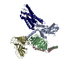
|
|---|---|
| 1 |
|
-要素
-Guanine nucleotide-binding protein ... , 3種, 3分子 ABC
| #2: タンパク質 | 分子量: 40446.047 Da / 分子数: 1 / 変異: G203A, A326S / 由来タイプ: 組換発現 / 由来: (組換発現) Homo sapiens (ヒト) / 遺伝子: GNAI1発現宿主:   Spodoptera frugiperda (ツマジロクサヨトウ) Spodoptera frugiperda (ツマジロクサヨトウ)参照: UniProt: P63096 |
|---|---|
| #4: タンパク質 | 分子量: 41772.562 Da / 分子数: 1 / 由来タイプ: 組換発現 / 由来: (組換発現) Mus musculus (ハツカネズミ) / 遺伝子: Gnb1発現宿主: Spodoptera frugiperda (ツマジロクサヨトウ)参照: UniProt: P62874 |
| #5: タンパク質 | 分子量: 7729.947 Da / 分子数: 1 / 由来タイプ: 組換発現 / 由来: (組換発現) Mus musculus (ハツカネズミ) / 遺伝子: Gng2発現宿主: Spodoptera frugiperda (ツマジロクサヨトウ)参照: UniProt: P63213 |
-タンパク質 / 抗体 / 非ポリマー , 3種, 3分子 RS
| #1: タンパク質 | 分子量: 60749.406 Da / 分子数: 1 / 変異: M29W,H124I / 由来タイプ: 組換発現 由来: (組換発現) Escherichia coli (大腸菌), (組換発現) Homo sapiens (ヒト)遺伝子: cybC, HCAR3, GPR109B, HCA3, HM74B, NIACR2 発現宿主: Spodoptera frugiperda (ツマジロクサヨトウ)参照: UniProt: P0ABE7, UniProt: P49019 |
|---|---|
| #3: 抗体 | 分子量: 26496.514 Da / 分子数: 1 / 由来タイプ: 組換発現 / 由来: (組換発現) synthetic construct (人工物) 発現宿主: Spodoptera frugiperda (ツマジロクサヨトウ) |
| #6: 化合物 | ChemComp-P9X / ( 分子量: 218.205 Da / 分子数: 1 / 由来タイプ: 合成 / 式: C12H10O4 / タイプ: SUBJECT OF INVESTIGATION 分子量: 218.205 Da / 分子数: 1 / 由来タイプ: 合成 / 式: C12H10O4 / タイプ: SUBJECT OF INVESTIGATION |
-詳細
| 研究の焦点であるリガンドがあるか | Y |
|---|
-実験情報
-実験
| 実験 | 手法: 電子顕微鏡法 |
|---|---|
| EM実験 | 試料の集合状態: PARTICLE / 3次元再構成法: 単粒子再構成法 |
- 試料調製
試料調製
| 構成要素 | 名称: Multiprotein complexタンパク質複合体 / タイプ: COMPLEX / Entity ID: #1-#2, #4-#5, #3 / 由来: MULTIPLE SOURCES |
|---|---|
| 分子量 | 実験値: NO |
| 由来(天然) | 生物種: Homo sapiens (ヒト) |
| 由来(組換発現) | 生物種: Spodoptera frugiperda (ツマジロクサヨトウ) |
| 緩衝液 | pH: 7.4 |
| 試料 | 濃度: 15 mg/ml / 包埋: NO / シャドウイング: NO / 染色: NO / 凍結: YES / 詳細: This sample was monodisperse |
| 急速凍結 | 装置: FEI VITROBOT MARK IV / 凍結剤: ETHANE / 湿度: 100 % / 凍結前の試料温度: 298 K |
- 電子顕微鏡撮影
| 顕微鏡 | モデル: JEOL CRYO ARM 300 |
|---|---|
| 電子銃 | 電子線源: FIELD EMISSION GUN / 加速電圧: 300 kV / 照射モード: FLOOD BEAM |
| 電子レンズ | モード: BRIGHT FIELDBright-field microscopy / 最大 デフォーカス(公称値): 2000 nm / 最小 デフォーカス(公称値): 1000 nm |
| 撮影 | 電子線照射量: 49 e/Å2 / フィルム・検出器のモデル: GATAN K3 (6k x 4k) |
- 解析
解析
| CTF補正 | タイプ: PHASE FLIPPING AND AMPLITUDE CORRECTION | ||||||||||||||||||||||||
|---|---|---|---|---|---|---|---|---|---|---|---|---|---|---|---|---|---|---|---|---|---|---|---|---|---|
| 3次元再構成 | 解像度: 3.07 Å / 解像度の算出法: FSC 0.143 CUT-OFF / 粒子像の数: 95567 / 対称性のタイプ: POINT | ||||||||||||||||||||||||
| 拘束条件 |
|
