 ムービー
ムービー コントローラー


+ データを開く

- 基本情報
基本情報
| 登録情報 | データベース: PDB / ID: 6eiw | |||||||||
|---|---|---|---|---|---|---|---|---|---|---|
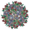
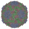
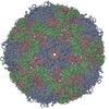
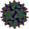
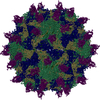
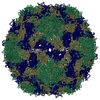
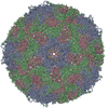
| タイトル | Sacbrood virus of honeybee empty particle | |||||||||
 要素 要素 |
| |||||||||
 キーワード キーワード |  VIRUS (ウイルス) / empty particle / icosahedral virus particle / iflavirus VIRUS (ウイルス) / empty particle / icosahedral virus particle / iflavirus | |||||||||
| 機能・相同性 |  機能・相同性情報 機能・相同性情報host cell membrane / カプシド / host cell cytoplasm / RNA helicase activity / viral RNA genome replication / cysteine-type endopeptidase activity / RNA-dependent RNA polymerase activity / DNA-templated transcription / structural molecule activity / タンパク質分解 ...host cell membrane / カプシド / host cell cytoplasm / RNA helicase activity / viral RNA genome replication / cysteine-type endopeptidase activity / RNA-dependent RNA polymerase activity / DNA-templated transcription / structural molecule activity / タンパク質分解 / RNA binding / ATP binding / 生体膜 / 細胞質類似検索 - 分子機能 | |||||||||
| 生物種 |  Sacbrood virus (ウイルス) Sacbrood virus (ウイルス) | |||||||||
| 手法 | 電子顕微鏡法 / 単粒子再構成法 / クライオ電子顕微鏡法 / 解像度: 3.87 Å | |||||||||
 データ登録者 データ登録者 | Plevka, P. / Prochazkova, M. | |||||||||
| 資金援助 |  チェコ, 2件 チェコ, 2件
| |||||||||
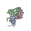
 引用 引用 | ジャーナル: Proc Natl Acad Sci U S A / 年: 2018 タイトル: Virion structure and genome delivery mechanism of sacbrood honeybee virus. 著者: Michaela Procházková / Tibor Füzik / Karel Škubník / Jana Moravcová / Zorica Ubiparip / Antonín Přidal / Pavel Plevka / 要旨: Infection by sacbrood virus (SBV) from the family Iflaviridae is lethal to honey bee larvae but only rarely causes the collapse of honey bee colonies. Despite the negative effect of SBV on honey ...Infection by sacbrood virus (SBV) from the family Iflaviridae is lethal to honey bee larvae but only rarely causes the collapse of honey bee colonies. Despite the negative effect of SBV on honey bees, the structure of its particles and mechanism of its genome delivery are unknown. Here we present the crystal structure of SBV virion and show that it contains 60 copies of a minor capsid protein (MiCP) attached to the virion surface. No similar MiCPs have been previously reported in any of the related viruses from the order Picornavirales. The location of the MiCP coding sequence within the SBV genome indicates that the MiCP evolved from a C-terminal extension of a major capsid protein by the introduction of a cleavage site for a virus protease. The exposure of SBV to acidic pH, which the virus likely encounters during cell entry, induces the formation of pores at threefold and fivefold axes of the capsid that are 7 Å and 12 Å in diameter, respectively. This is in contrast to vertebrate picornaviruses, in which the pores along twofold icosahedral symmetry axes are currently considered the most likely sites for genome release. SBV virions lack VP4 subunits that facilitate the genome delivery of many related dicistroviruses and picornaviruses. MiCP subunits induce liposome disruption in vitro, indicating that they are functional analogs of VP4 subunits and enable the virus genome to escape across the endosome membrane into the cell cytoplasm. | |||||||||
| 履歴 |
|
- 構造の表示
構造の表示
| ムービー |
ムービービューア |
|---|---|
| 構造ビューア | 分子: MolmilJmol/JSmol |
- ダウンロードとリンク
-ダウンロード
| PDBx/mmCIF形式 | 6eiw.cif.gz | 158.5 KB | 表示 | PDBx/mmCIF形式 |
|---|---|---|---|---|
| PDB形式 | pdb6eiw.ent.gz | 120.8 KB | 表示 | PDB形式 |
| PDBx/mmJSON形式 | 6eiw.json.gz | ツリー表示 | PDBx/mmJSON形式 | |
| その他 |  その他のダウンロード その他のダウンロード |
-検証レポート
| アーカイブディレクトリ | https://data.pdbj.org/pub/pdb/validation_reports/ei/6eiwftp://data.pdbj.org/pub/pdb/validation_reports/ei/6eiw | HTTPS FTP |
|---|
-関連構造データ
| 関連構造データ |  3881MC 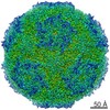 3863C 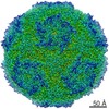 3865C  3866C  3867C 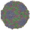 5lsfC  5oypC 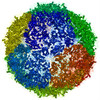 6egvC  6egxC  6eh1C M: このデータのモデリングに利用したマップデータ C: 同じ文献を引用 ( |
|---|---|
| 類似構造データ |

-リンク
 PDBj
PDBj
- 集合体
集合体
| 登録構造単位 | 
|
|---|---|
| 1 | x 60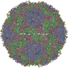
|
| 2 |
|
| 3 | x 5
|
| 4 | x 6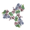
|
| 5 | 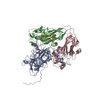
|
| 対称性 | 点対称性: (シェーンフリース記号: I (正20面体型対称)) |
-要素
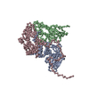
| #1: タンパク質 | 構造 分子量: 26151.500 Da / 分子数: 1 / 由来タイプ: 天然 / 由来: (天然) Sacbrood virus (ウイルス) / Plasmid details: Petrusov, CZ / 参照: UniProt: A0A223DN59, UniProt: Q9WCE9*PLUS |
|---|---|
| #2: タンパク質 | 構造 分子量: 22563.734 Da / 分子数: 1 / 由来タイプ: 天然 / 由来: (天然) Sacbrood virus (ウイルス) / Plasmid details: Petrusov, CZ / 参照: UniProt: A0A223DN66 |
| #3: タンパク質 | 構造 分子量: 30701.373 Da / 分子数: 1 / 由来タイプ: 天然 / 由来: (天然) Sacbrood virus (ウイルス) / Plasmid details: Petrusov, CZ / 参照: UniProt: A0A2I6HDZ6 |
| #4: タンパク質・ペプチド | 分子量: 3107.398 Da / 分子数: 1 / 由来タイプ: 天然 / 由来: (天然) Sacbrood virus (ウイルス) / Plasmid details: Petrusov, CZ / 参照: UniProt: Q9IGK7 |
-実験情報
-実験
| 実験 | 手法: 電子顕微鏡法 |
|---|---|
| EM実験 | 試料の集合状態: PARTICLE / 3次元再構成法: 単粒子再構成法 |
- 試料調製
試料調製
| 構成要素 | 名称: Sacbrood virus / タイプ: VIRUS / Entity ID: all / 由来: NATURAL | |||||||||||||||||||||||||
|---|---|---|---|---|---|---|---|---|---|---|---|---|---|---|---|---|---|---|---|---|---|---|---|---|---|---|
| 分子量 | 実験値: NO | |||||||||||||||||||||||||
| 由来(天然) | 生物種: Sacbrood virus (ウイルス) | |||||||||||||||||||||||||
| ウイルスについての詳細 | 中空か: YES / エンベロープを持つか: NO / 単離: STRAIN / タイプ: VIRION | |||||||||||||||||||||||||
| 天然宿主 | 生物種: Apis mellifera | |||||||||||||||||||||||||
| ウイルス殻 | 名称: capsidカプシド / 直径: 273.94 nm / 三角数 (T数): 3 | |||||||||||||||||||||||||
| 緩衝液 | pH: 7.4 | |||||||||||||||||||||||||
| 緩衝液成分 |
| |||||||||||||||||||||||||
| 試料 | 濃度: 15 mg/ml / 包埋: NO / シャドウイング: NO / 染色: NO / 凍結: YES / 詳細: empty particle | |||||||||||||||||||||||||
| 試料支持 | グリッドの材料: COPPER / グリッドのサイズ: 300 divisions/in. / グリッドのタイプ: Quantifoil R2/1 | |||||||||||||||||||||||||
| 急速凍結 | 装置: FEI VITROBOT MARK II / 凍結剤: ETHANE / 湿度: 70 % / 凍結前の試料温度: 298 K |
- 電子顕微鏡撮影
| 実験機器 |  モデル: Titan Krios / 画像提供: FEI Company |
|---|---|
| 顕微鏡 | モデル: FEI TITAN KRIOS 詳細: Preliminar grid screening was performed manually on FEI Tecnai F20 (cryo stage). |
| 電子銃 | 電子線源: FIELD EMISSION GUN / 加速電圧: 300 kV / 照射モード: FLOOD BEAM |
| 電子レンズ | モード: BRIGHT FIELDBright-field microscopy / 倍率(公称値): 75000 X / 倍率(補正後): 74325 X / 最大 デフォーカス(公称値): 4000 nm / 最小 デフォーカス(公称値): 1000 nm / Cs: 2.7 mm / C2レンズ絞り径: 100 µm / アライメント法: COMA FREE |
| 試料ホルダ | 凍結剤: NITROGEN 試料ホルダーモデル: FEI TITAN KRIOS AUTOGRID HOLDER |
| 撮影 | 平均露光時間: 1 sec. / 電子線照射量: 21 e/Å2 / 検出モード: COUNTING フィルム・検出器のモデル: FEI FALCON II (4k x 4k) 撮影したグリッド数: 1 |
| 画像スキャン | 横: 4096 / 縦: 4096 / 動画フレーム数/画像: 16 / 利用したフレーム数/画像: 2-16 |
- 解析
解析
| ソフトウェア | 名称: REFMAC / バージョン: 5.8.0222 / 分類: 精密化 | ||||||||||||||||||||||||||||||||||||||||||||||||||||||||||||||||||||||||||||||||||||||||||||||||||||||||||
|---|---|---|---|---|---|---|---|---|---|---|---|---|---|---|---|---|---|---|---|---|---|---|---|---|---|---|---|---|---|---|---|---|---|---|---|---|---|---|---|---|---|---|---|---|---|---|---|---|---|---|---|---|---|---|---|---|---|---|---|---|---|---|---|---|---|---|---|---|---|---|---|---|---|---|---|---|---|---|---|---|---|---|---|---|---|---|---|---|---|---|---|---|---|---|---|---|---|---|---|---|---|---|---|---|---|---|---|
| EMソフトウェア |
| ||||||||||||||||||||||||||||||||||||||||||||||||||||||||||||||||||||||||||||||||||||||||||||||||||||||||||
| CTF補正 | タイプ: PHASE FLIPPING AND AMPLITUDE CORRECTION | ||||||||||||||||||||||||||||||||||||||||||||||||||||||||||||||||||||||||||||||||||||||||||||||||||||||||||
| 粒子像の選択 | 選択した粒子像数: 31804 詳細: Full virions were selected together with empty particles from the same set of micrographs. Subsequent 2D classification sorted the particles of two types. Full and empty particles were ...詳細: Full virions were selected together with empty particles from the same set of micrographs. Subsequent 2D classification sorted the particles of two types. Full and empty particles were further reconstructed separately. | ||||||||||||||||||||||||||||||||||||||||||||||||||||||||||||||||||||||||||||||||||||||||||||||||||||||||||
| 3次元再構成 | 解像度: 3.87 Å / 解像度の算出法: FSC 0.143 CUT-OFF / 粒子像の数: 10303 / アルゴリズム: FOURIER SPACE / クラス平均像の数: 1 / 対称性のタイプ: POINT | ||||||||||||||||||||||||||||||||||||||||||||||||||||||||||||||||||||||||||||||||||||||||||||||||||||||||||
| 原子モデル構築 | B value: 99.36 / プロトコル: RIGID BODY FIT / 空間: REAL / Target criteria: R-factor | ||||||||||||||||||||||||||||||||||||||||||||||||||||||||||||||||||||||||||||||||||||||||||||||||||||||||||
| 精密化 | 解像度: 3.87→3.87 Å / Cor.coef. Fo:Fc: 0.919 / SU B: 21.779 / SU ML: 0.29 / ESU R: 0.147 立体化学のターゲット値: MAXIMUM LIKELIHOOD WITH PHASES 詳細: HYDROGENS HAVE BEEN ADDED IN THE RIDING POSITIONS
| ||||||||||||||||||||||||||||||||||||||||||||||||||||||||||||||||||||||||||||||||||||||||||||||||||||||||||
| 溶媒の処理 | イオンプローブ半径: 0.8 Å / 減衰半径: 0.8 Å / VDWプローブ半径: 1.2 Å / 溶媒モデル: MASK | ||||||||||||||||||||||||||||||||||||||||||||||||||||||||||||||||||||||||||||||||||||||||||||||||||||||||||
| 原子変位パラメータ | Biso mean: 88.209 Å2
| ||||||||||||||||||||||||||||||||||||||||||||||||||||||||||||||||||||||||||||||||||||||||||||||||||||||||||
| 精密化ステップ | サイクル: 1 / 合計: 5803 | ||||||||||||||||||||||||||||||||||||||||||||||||||||||||||||||||||||||||||||||||||||||||||||||||||||||||||
| 拘束条件 |
|