THE ENTRY PRESENTED HERE DOES NOT CONTAIN THE COMPLETE ASYMMETRIC UNIT. IT MAY BE GENERATED USING THE MTRIX TRANSFORMATIONS BELOW. ALSO SEE REMARKS 295 AND 300.
-
Components
#1: Protein
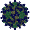
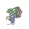
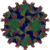
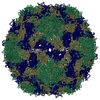
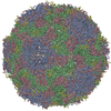
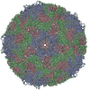
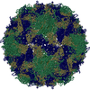
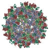
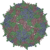
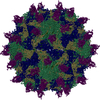
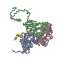
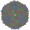
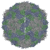
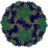
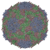
PROTEIN (CRICKETPARALYSISVIRUS, VP1) / CRPV
Mass: 28853.590 Da / Num. of mol.: 1 / Source method: isolated from a natural source / Source: (natural) Cricket paralysis virus / Genus: Cripavirus / References: UniProt: P13418
#2: Protein
PROTEIN (CRICKETPARALYSISVIRUS, VP2) / CRPV
Mass: 28709.201 Da / Num. of mol.: 1 / Source method: isolated from a natural source / Source: (natural) Cricket paralysis virus / Genus: Cripavirus / References: UniProt: P13418
#3: Protein
PROTEIN (CRICKETPARALYSISVIRUS, VP3) / CRPV
Mass: 31917.018 Da / Num. of mol.: 1 / Source method: isolated from a natural source / Source: (natural) Cricket paralysis virus / Genus: Cripavirus / References: UniProt: P13418
#4: Protein
PROTEIN (CRICKETPARALYSISVIRUS, VP4) / CRPV
Mass: 5932.688 Da / Num. of mol.: 1 / Source method: isolated from a natural source / Source: (natural) Cricket paralysis virus / Genus: Cripavirus / References: UniProt: P13418
Mass: 18.015 Da / Num. of mol.: 178 / Source method: isolated from a natural source / Formula: H2O
Compound details
A SALT BRIDGE IS VISIBLE IN ELECTRON DENSITY MAPS, BETWEEN RESIDUES ARG A 222 AND GLU A 112 FROM AN ...A SALT BRIDGE IS VISIBLE IN ELECTRON DENSITY MAPS, BETWEEN RESIDUES ARG A 222 AND GLU A 112 FROM AN ADJACENT, 5-FOLD RELATED COPY OF VP1.
-
Experimental details
-
Experiment
Experiment
Method: X-RAY DIFFRACTION / Number of used crystals: 1
-
Sample preparation
Crystal grow
pH: 6 Details: VIRUS STORED AT 10MG/ML IN 200MM NAH2PO4, PH7.2. WELL SOLUTION CONSISTED OF 8% (W/V) MPEG 5K, 50MM LI2SO4, 50MM MES, PH6.0.
Resolution: 2.4→15 Å / Rfactor Rfree error: 0.002 / Data cutoff high rms absF: 10511948.71 / Isotropic thermal model: RESTRAINED / σ(F): 2 Details: RESIDUES B 253 - B 271 ARE PRESENT IN THE MODEL, BUT THE SEQUENCE OF THESE RESIDUES DOES NOT CORRESPOND TO ANY PREDICTED SEQUENCE IN THE VIRAL GENOME. THEY ARE INCLUDED ONLY TO PARTIALLY ...Details: RESIDUES B 253 - B 271 ARE PRESENT IN THE MODEL, BUT THE SEQUENCE OF THESE RESIDUES DOES NOT CORRESPOND TO ANY PREDICTED SEQUENCE IN THE VIRAL GENOME. THEY ARE INCLUDED ONLY TO PARTIALLY MODEL THE POOR QUALITY, APPARENTLY HELICAL DENSITY ON THE VIRUS SURFACE AT THE CAPSID 2-FOLD AXES.
In the structure databanks used in Yorodumi, some data are registered as the other names, "COVID-19 virus" and "2019-nCoV". Here are the details of the virus and the list of structure data.
Jan 31, 2019. EMDB accession codes are about to change! (news from PDBe EMDB page)
EMDB accession codes are about to change! (news from PDBe EMDB page)
The allocation of 4 digits for EMDB accession codes will soon come to an end. Whilst these codes will remain in use, new EMDB accession codes will include an additional digit and will expand incrementally as the available range of codes is exhausted. The current 4-digit format prefixed with “EMD-” (i.e. EMD-XXXX) will advance to a 5-digit format (i.e. EMD-XXXXX), and so on. It is currently estimated that the 4-digit codes will be depleted around Spring 2019, at which point the 5-digit format will come into force.
The EM Navigator/Yorodumi systems omit the EMD- prefix.
Related info.:Q: What is EMD? / ID/Accession-code notation in Yorodumi/EM Navigator
Yorodumi is a browser for structure data from EMDB, PDB, SASBDB, etc.
This page is also the successor to EM Navigator detail page, and also detail information page/front-end page for Omokage search.
The word "yorodu" (or yorozu) is an old Japanese word meaning "ten thousand". "mi" (miru) is to see.
Related info.:EMDB / PDB / SASBDB / Comparison of 3 databanks / Yorodumi Search / Aug 31, 2016. New EM Navigator & Yorodumi / Yorodumi Papers / Jmol/JSmol / Function and homology information / Changes in new EM Navigator and Yorodumi
 Movie
Movie Controller


 Open data
 Basic information
 Components
 Keywords
 VIRUS / INSECT PICORNA-LIKE VIRUS / Icosahedral virus
 Function and homology information
 Authors
 Citation
 Structure visualization
 Downloads & links
 Other downloads
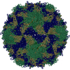
 PDBj
PDBj
 Assembly


 Mass: 18.015 Da / Num. of mol.: 178 / Source method: isolated from a natural source / Formula: H2O
 Sample preparation
 / Beamline: F1 / Wavelength: 0.928
  Processing