 ムービー
ムービー コントローラー


+ データを開く

- 基本情報
基本情報
| 登録情報 | データベース: PDB / ID: 5tat | ||||||
|---|---|---|---|---|---|---|---|
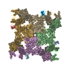
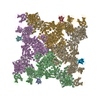
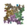
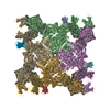
| タイトル | Structure of rabbit RyR1 (Caffeine/ATP/EGTA dataset, class 2) | ||||||
 要素 要素 |
| ||||||
 キーワード キーワード | TRANSPORT PROTEIN/ISOMERASE / RyR /  Ca2+ / EC coupling / gating / TRANSPORT PROTEIN-ISOMERASE complex Ca2+ / EC coupling / gating / TRANSPORT PROTEIN-ISOMERASE complex | ||||||
| 機能・相同性 |  機能・相同性情報 機能・相同性情報ATP-gated ion channel activity / positive regulation of sequestering of calcium ion / cyclic nucleotide binding / negative regulation of release of sequestered calcium ion into cytosol / terminal cisterna / negative regulation of insulin secretion involved in cellular response to glucose stimulus / ryanodine receptor complex / ryanodine-sensitive calcium-release channel activity / neuronal action potential propagation / insulin secretion involved in cellular response to glucose stimulus ...ATP-gated ion channel activity / positive regulation of sequestering of calcium ion / cyclic nucleotide binding / negative regulation of release of sequestered calcium ion into cytosol / terminal cisterna / negative regulation of insulin secretion involved in cellular response to glucose stimulus / ryanodine receptor complex / ryanodine-sensitive calcium-release channel activity / neuronal action potential propagation / insulin secretion involved in cellular response to glucose stimulus / release of sequestered calcium ion into cytosol by sarcoplasmic reticulum / ossification involved in bone maturation / cell communication by electrical coupling involved in cardiac conduction / response to redox state / protein maturation by protein folding / skin development / 'de novo' protein folding / negative regulation of heart rate / negative regulation of phosphoprotein phosphatase activity / FK506 binding / positive regulation of axon regeneration / cellular response to caffeine / intracellularly gated calcium channel activity / outflow tract morphogenesis / organelle membrane / : / toxic substance binding / smooth muscle contraction / negative regulation of ryanodine-sensitive calcium-release channel activity / voltage-gated calcium channel activity / response to vitamin E / calcium channel inhibitor activity / regulation of cardiac muscle contraction by regulation of the release of sequestered calcium ion / skeletal muscle fiber development / protein peptidyl-prolyl isomerization / T cell proliferation / regulation of release of sequestered calcium ion into cytosol by sarcoplasmic reticulum / release of sequestered calcium ion into cytosol / regulation of ryanodine-sensitive calcium-release channel activity / Ion homeostasis / sarcoplasmic reticulum membrane / calcium channel complex / regulation of cytosolic calcium ion concentration / cellular response to calcium ion / 筋小胞体 / muscle contraction / プロリルイソメラーゼ / peptidyl-prolyl cis-trans isomerase activity / calcium ion transmembrane transport / calcium channel activity / response to hydrogen peroxide / intracellular calcium ion homeostasis / Stimuli-sensing channels / Z disc / disordered domain specific binding / positive regulation of cytosolic calcium ion concentration / protein refolding / protein homotetramerization / transmembrane transporter binding / calmodulin binding / signaling receptor binding / calcium ion binding / ATP binding / 生体膜 / identical protein binding / 細胞質基質 / 細胞質類似検索 - 分子機能 | ||||||
| 生物種 |  Homo sapiens (ヒト) Homo sapiens (ヒト) Oryctolagus cuniculus (ウサギ) Oryctolagus cuniculus (ウサギ) | ||||||
| 手法 | 電子顕微鏡法 / 単粒子再構成法 / クライオ電子顕微鏡法 / 解像度: 4.8 Å | ||||||
 データ登録者 データ登録者 | Clarke, O.B. / des Georges, A. / Zalk, R. / Marks, A.R. / Hendrickson, W.A. / Frank, J. | ||||||
 引用 引用 | ジャーナル: Cell / 年: 2016 タイトル: Structural Basis for Gating and Activation of RyR1. 著者: Amédée des Georges / Oliver B Clarke / Ran Zalk / Qi Yuan / Kendall J Condon / Robert A Grassucci / Wayne A Hendrickson / Andrew R Marks / Joachim Frank /  要旨: The type-1 ryanodine receptor (RyR1) is an intracellular calcium (Ca(2+)) release channel required for skeletal muscle contraction. Here, we present cryo-EM reconstructions of RyR1 in multiple ...The type-1 ryanodine receptor (RyR1) is an intracellular calcium (Ca(2+)) release channel required for skeletal muscle contraction. Here, we present cryo-EM reconstructions of RyR1 in multiple functional states revealing the structural basis of channel gating and ligand-dependent activation. Binding sites for the channel activators Ca(2+), ATP, and caffeine were identified at interdomain interfaces of the C-terminal domain. Either ATP or Ca(2+) alone induces conformational changes in the cytoplasmic assembly ("priming"), without pore dilation. In contrast, in the presence of all three activating ligands, high-resolution reconstructions of open and closed states of RyR1 were obtained from the same sample, enabling analyses of conformational changes associated with gating. Gating involves global conformational changes in the cytosolic assembly accompanied by local changes in the transmembrane domain, which include bending of the S6 transmembrane segment and consequent pore dilation, displacement, and deformation of the S4-S5 linker and conformational changes in the pseudo-voltage-sensor domain. | ||||||
| 履歴 |
|
- 構造の表示
構造の表示
| ムービー |
ムービービューア |
|---|---|
| 構造ビューア | 分子: MolmilJmol/JSmol |
- ダウンロードとリンク
-ダウンロード
| PDBx/mmCIF形式 | 5tat.cif.gz | 2.7 MB | 表示 | PDBx/mmCIF形式 |
|---|---|---|---|---|
| PDB形式 | pdb5tat.ent.gz | 表示 | PDB形式 | |
| PDBx/mmJSON形式 | 5tat.json.gz | ツリー表示 | PDBx/mmJSON形式 | |
| その他 |  その他のダウンロード その他のダウンロード |
-検証レポート
| アーカイブディレクトリ | https://data.pdbj.org/pub/pdb/validation_reports/ta/5tatftp://data.pdbj.org/pub/pdb/validation_reports/ta/5tat | HTTPS FTP |
|---|
-関連構造データ
| 関連構造データ | 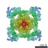 8384MC 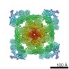 8342C 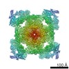 8372C 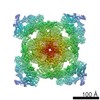 8373C 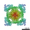 8374C 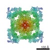 8375C 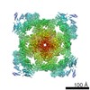 8376C 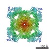 8377C 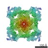 8378C 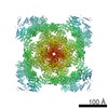 8379C 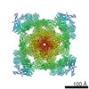 8380C 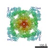 8381C 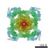 8382C 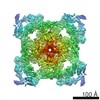 8383C 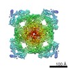 8385C 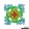 8386C 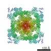 8387C 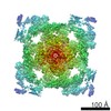 8388C 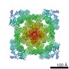 8389C 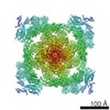 8390C 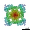 8391C 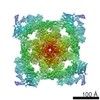 8392C 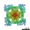 8393C 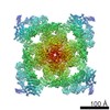 8394C 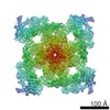 8395C  5t15C  5t9mC  5t9nC  5t9rC  5t9sC  5t9vC  5ta3C  5talC  5tamC  5tanC  5tapC  5taqC  5tasC  5tauC  5tavC  5tawC  5taxC  5tayC  5tazC  5tb0C  5tb1C  5tb2C  5tb3C  5tb4C M: このデータのモデリングに利用したマップデータ C: 同じ文献を引用 ( |
|---|---|
| 類似構造データ |

-リンク
 PDBj
PDBj


- 集合体
集合体
| 登録構造単位 | 
|
|---|---|
| 1 |
|
-要素
| #1: タンパク質 | 分子量: 11798.501 Da / 分子数: 4 / 由来タイプ: 組換発現 / 由来: (組換発現) Homo sapiens (ヒト) / 遺伝子: FKBP1B, FKBP12.6, FKBP1L, FKBP9, OTK4 / 発現宿主:  Escherichia coli (大腸菌) / 参照: UniProt: P68106, プロリルイソメラーゼ Escherichia coli (大腸菌) / 参照: UniProt: P68106, プロリルイソメラーゼ#2: タンパク質 | / RyR1 / Skeletal muscle calcium release channel / Skeletal muscle ryanodine receptor / Skeletal ...RyR1 / Skeletal muscle calcium release channel / Skeletal muscle ryanodine receptor / Skeletal muscle-type ryanodine receptor / Type 1 ryanodine receptor分子量: 475107.719 Da / 分子数: 4 / 由来タイプ: 天然 / 由来: (天然) Oryctolagus cuniculus (ウサギ) / 組織: Skeletal muscle骨格筋 / 参照: UniProt: P11716#3: 化合物 | ChemComp-ATP / アデノシン三リン酸  分子量: 507.181 Da / 分子数: 4 / 由来タイプ: 合成 / 式: C10H16N5O13P3 / コメント: ATP, エネルギー貯蔵分子*YM 分子量: 507.181 Da / 分子数: 4 / 由来タイプ: 合成 / 式: C10H16N5O13P3 / コメント: ATP, エネルギー貯蔵分子*YM#4: 化合物 | ChemComp-CFF / Caffeine (data page)  分子量: 194.191 Da / 分子数: 4 / 由来タイプ: 合成 / 式: C8H10N4O2 / コメント: 薬剤*YM 分子量: 194.191 Da / 分子数: 4 / 由来タイプ: 合成 / 式: C8H10N4O2 / コメント: 薬剤*YM#5: 化合物 | ChemComp-ZN /   分子量: 65.409 Da / 分子数: 4 / 由来タイプ: 合成 / 式: Zn 分子量: 65.409 Da / 分子数: 4 / 由来タイプ: 合成 / 式: Zn |
|---|
-実験情報
-実験
| 実験 | 手法: 電子顕微鏡法 |
|---|---|
| EM実験 | 試料の集合状態: PARTICLE / 3次元再構成法: 単粒子再構成法 |
- 試料調製
試料調製
| 構成要素 | 名称: RyR1-Cs2 complex / タイプ: COMPLEX / Entity ID: #1-#2 / 由来: MULTIPLE SOURCES |
|---|---|
| 緩衝液 | pH: 7.4 |
| 試料 | 濃度: 6 mg/ml / 包埋: NO / シャドウイング: NO / 染色: NO / 凍結: YES |
| 試料支持 | グリッドの材料: GOLD / グリッドのサイズ: 400 divisions/in. / グリッドのタイプ: Quantifoil |
| 急速凍結 | 装置: FEI VITROBOT MARK IV / 凍結剤: ETHANE / 湿度: 100 % / 凍結前の試料温度: 277 K 詳細: Blotted for 3-4 seconds on both sides with Whatman ashless filter paper, blot force 3, wait time 30 seconds |
- 電子顕微鏡撮影
| 実験機器 |  モデル: Tecnai Polara / 画像提供: FEI Company |
|---|---|
| 顕微鏡 | モデル: FEI POLARA 300 |
| 電子銃 | 電子線源: FIELD EMISSION GUN / 加速電圧: 300 kV / 照射モード: FLOOD BEAM |
| 電子レンズ | モード: BRIGHT FIELDBright-field microscopy |
| 撮影 | 電子線照射量: 50 e/Å2 / 検出モード: COUNTING フィルム・検出器のモデル: GATAN K2 SUMMIT (4k x 4k) |
- 解析
解析
| EMソフトウェア |
| ||||||||||||||||||||||||||||||||||||
|---|---|---|---|---|---|---|---|---|---|---|---|---|---|---|---|---|---|---|---|---|---|---|---|---|---|---|---|---|---|---|---|---|---|---|---|---|---|
| CTF補正 | タイプ: PHASE FLIPPING AND AMPLITUDE CORRECTION | ||||||||||||||||||||||||||||||||||||
| 3次元再構成 | 解像度: 4.8 Å / 解像度の算出法: FSC 0.143 CUT-OFF / 粒子像の数: 55564 / アルゴリズム: FOURIER SPACE 詳細: The reported resolution is for the core. The resolution of the whole assembly is 5.3 Angstrom. 対称性のタイプ: POINT |
