 Movie
Movie Controller

[English] 日本語
 Yorodumi
- PDB-6fg3: Structure of Ryanodine receptor 1 in nanodiscs in the presence of... -
+ Open data

- Basic information
| Entry | Database: PDB / ID: 6fg3 | |||||||||
|---|---|---|---|---|---|---|---|---|---|---|
| Title | Structure of Ryanodine receptor 1 in nanodiscs in the presence of calcium, ATP and ryanodine | |||||||||
 Components Components | Ryanodine receptor 1 | |||||||||
 Keywords Keywords | MEMBRANE PROTEIN / calcium release channel / nanodiscs | |||||||||
| Function / homology |  Function and homology information Function and homology informationATP-gated ion channel activity / terminal cisterna / ryanodine receptor complex / ryanodine-sensitive calcium-release channel activity / release of sequestered calcium ion into cytosol by sarcoplasmic reticulum / ossification involved in bone maturation / skin development / cellular response to caffeine / intracellularly gated calcium channel activity / outflow tract morphogenesis ...ATP-gated ion channel activity / terminal cisterna / ryanodine receptor complex / ryanodine-sensitive calcium-release channel activity / release of sequestered calcium ion into cytosol by sarcoplasmic reticulum / ossification involved in bone maturation / skin development / cellular response to caffeine / intracellularly gated calcium channel activity / outflow tract morphogenesis / organelle membrane / toxic substance binding / voltage-gated calcium channel activity / skeletal muscle fiber development / release of sequestered calcium ion into cytosol / sarcoplasmic reticulum membrane / cellular response to calcium ion / sarcoplasmic reticulum / muscle contraction / calcium ion transmembrane transport / calcium channel activity / intracellular calcium ion homeostasis / disordered domain specific binding / protein homotetramerization / transmembrane transporter binding / calmodulin binding / calcium ion binding / ATP binding / membrane / identical protein bindingSimilarity search - Function | |||||||||
| Biological species |  Oryctolagus cuniculus (rabbit) Oryctolagus cuniculus (rabbit) | |||||||||
| Method | ELECTRON MICROSCOPY / single particle reconstruction / cryo EM / Resolution: 7.3 Å | |||||||||
 Authors Authors | Willegems, K. / Efremov, R.G. | |||||||||
| Funding support |  Belgium, 2items Belgium, 2items
| |||||||||
 Citation Citation | Journal: Structure / Year: 2018 Title: Influence of Lipid Mimetics on Gating of Ryanodine Receptor. Authors: Katrien Willegems / Rouslan G Efremov / Abstract: Understanding gating principles of ion channels at high resolution is of great importance. Here we investigate the conformational transition from closed to open state in ryanodine receptor 1 (RyR1) ...Understanding gating principles of ion channels at high resolution is of great importance. Here we investigate the conformational transition from closed to open state in ryanodine receptor 1 (RyR1) reconstituted into lipid nanodiscs. RyR1 is a homotetrameric giant ion channel that couples excitation of muscle cells to fast calcium release from the sarcoplasmic reticulum. Using single-particle cryo-EM we show that RyR1 reconstituted into lipid nanodiscs is stabilized in the open conformation when bound to the plant toxin ryanodine, but not in the presence of its physiological activators, calcium and ATP. Further, using ryanodine binding assays we show that membrane mimetics influence RyR1 transition between closed and open-channel conformations. We find that all detergents, including fluorinated detergents added to nanodiscs, stabilize closed state of RyR1. Our biochemical results correlate with available structural data and suggest optimal conditions for structural studies of RyR1 gating. | |||||||||
| History |
|
- Structure visualization
| Movie |
Movie viewer |
|---|---|
| Structure viewer | Molecule: MolmilJmol/JSmol |
- Downloads & links
-Download
| PDBx/mmCIF format | 6fg3.cif.gz | 2.6 MB | Display | PDBx/mmCIF format |
|---|---|---|---|---|
| PDB format | pdb6fg3.ent.gz | Display | PDB format | |
| PDBx/mmJSON format | 6fg3.json.gz | Tree view | PDBx/mmJSON format | |
| Others |  Other downloads Other downloads |
-Validation report
| Arichive directory | https://data.pdbj.org/pub/pdb/validation_reports/fg/6fg3ftp://data.pdbj.org/pub/pdb/validation_reports/fg/6fg3 | HTTPS FTP |
|---|
-Related structure data
| Related structure data | 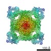 4258MC 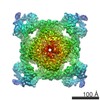 4295C  6fooC M: map data used to model this data C: citing same article ( |
|---|---|
| Similar structure data |

-Links
 PDBj
PDBj

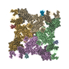
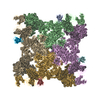
- Assembly
| Deposited unit | 
|
|---|---|
| 1 |
|
-Components
| #1: Protein | / RyR1 / Skeletal muscle calcium release channel / Skeletal muscle ryanodine receptor / Skeletal ...RyR1 / Skeletal muscle calcium release channel / Skeletal muscle ryanodine receptor / Skeletal muscle-type ryanodine receptor / Type 1 ryanodine receptor Mass: 532033.062 Da / Num. of mol.: 4 / Source method: isolated from a natural source Details: Poly Unk stretches correspond to polypeptide regions where the register is not assigned. Source: (natural) Oryctolagus cuniculus (rabbit) / Strain: New Zealand white rabbit / Tissue: skeletal muscle / References: UniProt: P11716#2: Chemical | ChemComp-ZN /   Mass: 65.409 Da / Num. of mol.: 4 / Source method: obtained synthetically / Formula: Zn Mass: 65.409 Da / Num. of mol.: 4 / Source method: obtained synthetically / Formula: Zn#3: Chemical | ChemComp-CA /   Mass: 40.078 Da / Num. of mol.: 4 / Source method: obtained synthetically / Formula: Ca Mass: 40.078 Da / Num. of mol.: 4 / Source method: obtained synthetically / Formula: Ca |
|---|
-Experimental details
-Experiment
| Experiment | Method: ELECTRON MICROSCOPY |
|---|---|
| EM experiment | Aggregation state: PARTICLE / 3D reconstruction method: single particle reconstruction |
- Sample preparation
| Component | Name: Ryanodine receptor 1 reconstituted in lipid nanodiscs in the presence of calcium, ATP and ryanodine Type: COMPLEX / Entity ID: #1 / Source: NATURAL | |||||||||||||||||||||||||||||||||||||||||||||
|---|---|---|---|---|---|---|---|---|---|---|---|---|---|---|---|---|---|---|---|---|---|---|---|---|---|---|---|---|---|---|---|---|---|---|---|---|---|---|---|---|---|---|---|---|---|---|
| Molecular weight | Value: 2.2 MDa / Experimental value: NO | |||||||||||||||||||||||||||||||||||||||||||||
| Source (natural) | Organism: Oryctolagus cuniculus (rabbit) / Strain: New Zealand white rabbit / Cellular location: sarcoplasmic reticulum / Tissue: skeletal muscle | |||||||||||||||||||||||||||||||||||||||||||||
| Buffer solution | pH: 7.4 | |||||||||||||||||||||||||||||||||||||||||||||
| Buffer component |
| |||||||||||||||||||||||||||||||||||||||||||||
| Specimen | Conc.: 3 mg/ml / Embedding applied: NO / Shadowing applied: NO / Staining applied: NO / Vitrification applied: YES | |||||||||||||||||||||||||||||||||||||||||||||
| Specimen support | Details: Glow discharged in ELMO plasma cleaner at current of 3 mA for 1 min. Grid material: COPPER / Grid mesh size: 300 divisions/in. / Grid type: Quantifoil R2/1 | |||||||||||||||||||||||||||||||||||||||||||||
| Vitrification | Instrument: HOMEMADE PLUNGER / Cryogen name: ETHANE / Chamber temperature: 293.15 K |
- Electron microscopy imaging
| Experimental equipment |  Model: Titan Krios / Image courtesy: FEI Company |
|---|---|
| Microscopy | Model: FEI TITAN KRIOS |
| Electron gun | Electron source: FIELD EMISSION GUN / Accelerating voltage: 300 kV / Illumination mode: FLOOD BEAM |
| Electron lens | Mode: BRIGHT FIELDBright-field microscopy / Nominal magnification: 59000 X / Nominal defocus max: 3000 nm / Nominal defocus min: 2000 nm |
| Specimen holder | Cryogen: NITROGEN |
| Image recording | Average exposure time: 1 sec. / Electron dose: 28 e/Å2 / Detector mode: COUNTING / Film or detector model: FEI FALCON II (4k x 4k) / Num. of grids imaged: 1 / Num. of real images: 3078 |
| Image scans | Sampling size: 14 µm / Width: 4096 / Height: 4096 / Movie frames/image: 7 / Used frames/image: 1-7 |
- Processing
| Software |
| ||||||||||||||||||||||||||||||||||||
|---|---|---|---|---|---|---|---|---|---|---|---|---|---|---|---|---|---|---|---|---|---|---|---|---|---|---|---|---|---|---|---|---|---|---|---|---|---|
| EM software |
| ||||||||||||||||||||||||||||||||||||
| CTF correction | Type: PHASE FLIPPING AND AMPLITUDE CORRECTION | ||||||||||||||||||||||||||||||||||||
| Particle selection | Num. of particles selected: 75794 | ||||||||||||||||||||||||||||||||||||
| Symmetry | Point symmetry: C4 (4 fold cyclic) | ||||||||||||||||||||||||||||||||||||
| 3D reconstruction | Resolution: 7.3 Å / Resolution method: FSC 0.143 CUT-OFF / Num. of particles: 61773 / Symmetry type: POINT | ||||||||||||||||||||||||||||||||||||
| Atomic model building | Protocol: FLEXIBLE FIT / Space: REAL | ||||||||||||||||||||||||||||||||||||
| Refinement | Stereochemistry target values: GeoStd + Monomer Library | ||||||||||||||||||||||||||||||||||||
| Refine LS restraints |
|
