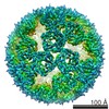
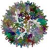
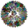
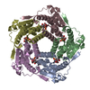
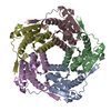
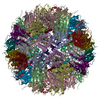
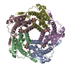
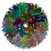
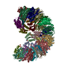
ジャーナル: Nat Commun / 年: 2017 タイトル: Structure and assembly of scalable porous protein cages. 著者: Eita Sasaki / Daniel Böhringer / Michiel van de Waterbeemd / Marc Leibundgut / Reinhard Zschoche / Albert J R Heck / Nenad Ban / Donald Hilvert / 要旨: Proteins that self-assemble into regular shell-like polyhedra are useful, both in nature and in the laboratory, as molecular containers. Here we describe cryo-electron microscopy (EM) structures of ...Proteins that self-assemble into regular shell-like polyhedra are useful, both in nature and in the laboratory, as molecular containers. Here we describe cryo-electron microscopy (EM) structures of two versatile encapsulation systems that exploit engineered electrostatic interactions for cargo loading. We show that increasing the number of negative charges on the lumenal surface of lumazine synthase, a protein that naturally assembles into a ∼1-MDa dodecahedron composed of 12 pentamers, induces stepwise expansion of the native protein shell, giving rise to thermostable ∼3-MDa and ∼6-MDa assemblies containing 180 and 360 subunits, respectively. Remarkably, these expanded particles assume unprecedented tetrahedrally and icosahedrally symmetric structures constructed entirely from pentameric units. Large keyhole-shaped pores in the shell, not present in the wild-type capsid, enable diffusion-limited encapsulation of complementarily charged guests. The structures of these supercharged assemblies demonstrate how programmed electrostatic effects can be effectively harnessed to tailor the architecture and properties of protein cages.
履歴
登録
2016年12月17日
登録サイト: PDBE / 処理サイト: PDBE
改定 1.0
2017年3月22日
Provider: repository / タイプ: Initial release
改定 1.1
2018年10月17日
Group: Data collection / カテゴリ: em_image_scans
改定 1.2
2019年7月31日
Group: Data collection / Refinement description / カテゴリ: refine
 ムービー
ムービー コントローラー


 データを開く
 基本情報
基本情報 要素
要素 Lumazine synthase
  キーワード
キーワード 機能・相同性情報

 データ登録者
 スイス, 1件
  引用
引用
 構造の表示
構造の表示 ダウンロードとリンク
 その他のダウンロード


 PDBj
PDBj
 集合体
集合体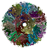
 試料調製
試料調製 電子顕微鏡撮影
 解析
解析