 Movie
Movie Controller

[English] 日本語
 Yorodumi
- PDB-1nqv: Crystal Structure of Lumazine Synthase from Aquifex aeolicus in C... -
+ Open data

- Basic information
| Entry | Database: PDB / ID: 1nqv | ||||||
|---|---|---|---|---|---|---|---|
| Title | Crystal Structure of Lumazine Synthase from Aquifex aeolicus in Complex with Inhibitor: 5-nitroso-6-ribityl-amino-2,4(1H,3H)pyrimidinedione | ||||||
 Components Components | 6,7-dimethyl-8-ribityllumazine synthase Lumazine synthase Lumazine synthase | ||||||
 Keywords Keywords | TRANSFERASE / Lumazine synthase / Aquifex aeolicus / inhibitor complex / vitamin biosynthesis / catalytic mechanism | ||||||
| Function / homology |  Function and homology information6,7-dimethyl-8-ribityllumazine synthase / 6,7-dimethyl-8-ribityllumazine synthase activity / riboflavin synthase complex / riboflavin biosynthetic process / cytosol Function and homology information6,7-dimethyl-8-ribityllumazine synthase / 6,7-dimethyl-8-ribityllumazine synthase activity / riboflavin synthase complex / riboflavin biosynthetic process / cytosolSimilarity search - Function | ||||||
| Biological species |   Aquifex aeolicus (bacteria) Aquifex aeolicus (bacteria) | ||||||
| Method | X-RAY DIFFRACTION / SYNCHROTRON / MOLECULAR REPLACEMENT / Resolution: 2.05 Å | ||||||
 Authors Authors | Zhang, X. / Meining, W. / Cushman, M. / Haase, I. / Fischer, M. / Bacher, A. / Ladenstein, R. | ||||||
 Citation Citation | Journal: J.Mol.Biol. / Year: 2003 Title: A structure-based model of the reaction catalyzed by lumazine synthase from Aquifex aeolicus. Authors: Zhang, X. / Meining, W. / Cushman, M. / Haase, I. / Fischer, M. / Bacher, A. / Ladenstein, R. | ||||||
| History |
|
- Structure visualization
| Structure viewer | Molecule: MolmilJmol/JSmol |
|---|
- Downloads & links
-Download
| PDBx/mmCIF format | 1nqv.cif.gz | 166.8 KB | Display | PDBx/mmCIF format |
|---|---|---|---|---|
| PDB format | pdb1nqv.ent.gz | 134.7 KB | Display | PDB format |
| PDBx/mmJSON format | 1nqv.json.gz | Tree view | PDBx/mmJSON format | |
| Others |  Other downloads Other downloads |
-Validation report
| Arichive directory | https://data.pdbj.org/pub/pdb/validation_reports/nq/1nqvftp://data.pdbj.org/pub/pdb/validation_reports/nq/1nqv | HTTPS FTP |
|---|
-Related structure data


-Links
 PDBj
PDBj- Assembly
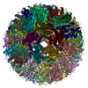
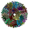
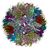
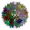
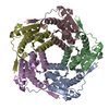
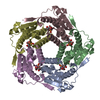
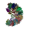
| Deposited unit | 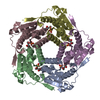
| |||||||||
|---|---|---|---|---|---|---|---|---|---|---|
| 1 | x 12
| |||||||||
| Unit cell |
| |||||||||
| Components on special symmetry positions |
| |||||||||
| Details | The biological assembly is an icosahedral capsid generated from the pentamer in the asymmetric unit by the I23 crystllographic symmetry operactions |
-Components
| #1: Protein | Lumazine synthase / DMRL synthase / Lumazine synthase / Riboflavin synthase beta chain Mass: 16727.201 Da / Num. of mol.: 5 Source method: isolated from a genetically manipulated source Source: (gene. exp.) Aquifex aeolicus (bacteria) / Production host: Escherichia coli (E. coli)References: UniProt: O66529, 6,7-dimethyl-8-ribityllumazine synthase#2: Chemical | ChemComp-PO4 / Phosphate  Mass: 94.971 Da / Num. of mol.: 5 / Source method: obtained synthetically / Formula: PO4 Mass: 94.971 Da / Num. of mol.: 5 / Source method: obtained synthetically / Formula: PO4#3: Chemical | ChemComp-LMZ /   Mass: 290.230 Da / Num. of mol.: 5 / Source method: obtained synthetically / Formula: C9H14N4O7 Mass: 290.230 Da / Num. of mol.: 5 / Source method: obtained synthetically / Formula: C9H14N4O7#4: Water | ChemComp-HOH / | Water Mass: 18.015 Da / Num. of mol.: 515 / Source method: isolated from a natural source / Formula: H2O Mass: 18.015 Da / Num. of mol.: 515 / Source method: isolated from a natural source / Formula: H2O |
|---|
-Experimental details
-Experiment
| Experiment | Method: X-RAY DIFFRACTION / Number of used crystals: 1 |
|---|
- Sample preparation
| Crystal | Density Matthews: 2.91 Å3/Da / Density % sol: 57.74 % | ||||||||||||||||||||||||||||||
|---|---|---|---|---|---|---|---|---|---|---|---|---|---|---|---|---|---|---|---|---|---|---|---|---|---|---|---|---|---|---|---|
| Crystal grow | Temperature: 293 K / Method: vapor diffusion, sitting drop / pH: 7.5 Details: sodium-potassium tartrate, HEPES, pH 7.5, VAPOR DIFFUSION, SITTING DROP, temperature 293.0K | ||||||||||||||||||||||||||||||
| Crystal grow | *PLUS pH: 7 / Method: vapor diffusion, sitting drop | ||||||||||||||||||||||||||||||
| Components of the solutions | *PLUS
|
-Data collection
| Diffraction | Mean temperature: 100 K |
|---|---|
| Diffraction source | Source: SYNCHROTRON / Site: EMBL/DESY, HAMBURG  / Beamline: X11 / Wavelength: 0.8482 Å / Beamline: X11 / Wavelength: 0.8482 Å |
| Detector | Type: MARRESEARCH / Detector: CCD / Date: Jun 8, 2001 |
| Radiation | Protocol: SINGLE WAVELENGTH / Monochromatic (M) / Laue (L): M / Scattering type: x-ray |
| Radiation wavelength | Wavelength: 0.8482 Å / Relative weight: 1 |
| Reflection | Resolution: 2.05→48.14 Å / Num. all: 60719 / Num. obs: 60309 / % possible obs: 99.3 % / Observed criterion σ(F): 0 / Observed criterion σ(I): 0 |
| Reflection shell | Resolution: 2.05→2.09 Å / % possible all: 100 |
| Reflection | *PLUS Num. obs: 60538 / % possible obs: 99.4 % / Num. measured all: 480544 / Rmerge(I) obs: 0.087 |
| Reflection shell | *PLUS Rmerge(I) obs: 0.297 / Mean I/σ(I) obs: 5 |
- Processing
| Software |
| ||||||||||||||||||||||||
|---|---|---|---|---|---|---|---|---|---|---|---|---|---|---|---|---|---|---|---|---|---|---|---|---|---|
| Refinement | Method to determine structure: MOLECULAR REPLACEMENT / Resolution: 2.05→48.14 Å / σ(F): 0 / Stereochemistry target values: Engh & Huber
| ||||||||||||||||||||||||
| Refinement step | Cycle: LAST / Resolution: 2.05→48.14 Å
| ||||||||||||||||||||||||
| Software | *PLUS Version: 5 / Classification: refinement | ||||||||||||||||||||||||
| Refine LS restraints | *PLUS
|
