 ムービー
ムービー コントローラー


+ データを開く

- 基本情報
基本情報
| 登録情報 | データベース: PDB / ID: 3dny | ||||||
|---|---|---|---|---|---|---|---|
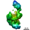
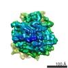
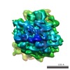
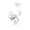
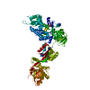
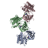
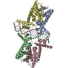
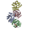
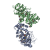
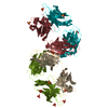
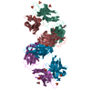
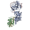
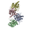
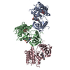
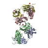
| タイトル | Fitting of the eEF2 crystal structure into the cryo-EM density map of the eEF2.80S.AlF4-.GDP complex | ||||||
 要素 要素 | Elongation factor 2 | ||||||
 キーワード キーワード | CELL CYCLE / eEF2 transition state complex / 80S Ribosome / AlF4- / GDP / GTPase / Translocation / Elongation factor / GTP-binding / Nucleotide-binding / Phosphoprotein / Protein biosynthesis / RNA-binding / rRNA-binding | ||||||
| 機能・相同性 |  機能・相同性情報 機能・相同性情報Peptide chain elongation / Synthesis of diphthamide-EEF2 / positive regulation of translational elongation / Protein methylation / translational elongation / translation elongation factor activity / Neutrophil degranulation / 加水分解酵素; 酸無水物に作用; GTPに作用・細胞または細胞小器官の運動に関与 / maintenance of translational fidelity / ribosome binding ...Peptide chain elongation / Synthesis of diphthamide-EEF2 / positive regulation of translational elongation / Protein methylation / translational elongation / translation elongation factor activity / Neutrophil degranulation / 加水分解酵素; 酸無水物に作用; GTPに作用・細胞または細胞小器官の運動に関与 / maintenance of translational fidelity / ribosome binding / protein-folding chaperone binding / rRNA binding / ribonucleoprotein complex / GTPase activity / GTP binding / identical protein binding / cytosol 類似検索 - 分子機能 | ||||||
| 生物種 |  | ||||||
| 手法 | 電子顕微鏡法 / 単粒子再構成法 / クライオ電子顕微鏡法 / 解像度: 12.6 Å | ||||||
 データ登録者 データ登録者 | Sengupta, J. / Frank, J. | ||||||
 引用 引用 | ジャーナル: J Mol Biol / 年: 2008 タイトル: Visualization of the eEF2-80S ribosome transition-state complex by cryo-electron microscopy. 著者: Jayati Sengupta / Jakob Nilsson / Richard Gursky / Morten Kjeldgaard / Poul Nissen / Joachim Frank /  要旨: In an attempt to understand ribosome-induced GTP hydrolysis on eEF2, we determined a 12.6-A cryo-electron microscopy reconstruction of the eEF2-bound 80S ribosome in the presence of aluminum ...In an attempt to understand ribosome-induced GTP hydrolysis on eEF2, we determined a 12.6-A cryo-electron microscopy reconstruction of the eEF2-bound 80S ribosome in the presence of aluminum tetrafluoride and GDP, with aluminum tetrafluoride mimicking the gamma-phosphate during hydrolysis. This is the first visualization of a structure representing a transition-state complex on the ribosome. Tight interactions are observed between the factor's G domain and the large ribosomal subunit, as well as between domain IV and an intersubunit bridge. In contrast, some of the domains of eEF2 implicated in small subunit binding display a large degree of flexibility. Furthermore, we find support for a transition-state model conformation of the switch I region in this complex where the reoriented switch I region interacts with a conserved rRNA region of the 40S subunit formed by loops of the 18S RNA helices 8 and 14. This complex is structurally distinct from the eEF2-bound 80S ribosome complexes previously reported, and analysis of this map sheds light on the GTPase-coupled translocation mechanism. | ||||||
| 履歴 |
|
- 構造の表示
構造の表示
| ムービー |
ムービービューア |
|---|---|
| 構造ビューア | 分子: MolmilJmol/JSmol |
- ダウンロードとリンク
-ダウンロード
| PDBx/mmCIF形式 | 3dny.cif.gz | 38.2 KB | 表示 | PDBx/mmCIF形式 |
|---|---|---|---|---|
| PDB形式 | pdb3dny.ent.gz | 19 KB | 表示 | PDB形式 |
| PDBx/mmJSON形式 | 3dny.json.gz | ツリー表示 | PDBx/mmJSON形式 | |
| その他 |  その他のダウンロード その他のダウンロード |
-検証レポート
| 文書・要旨 | 3dny_validation.pdf.gz | 549.6 KB | 表示 | wwPDB検証レポート |
|---|---|---|---|---|
| 文書・詳細版 | 3dny_full_validation.pdf.gz | 549.2 KB | 表示 | |
| XML形式データ | 3dny_validation.xml.gz | 14 KB | 表示 | |
| CIF形式データ | 3dny_validation.cif.gz | 19.7 KB | 表示 | |
| アーカイブディレクトリ | https://data.pdbj.org/pub/pdb/validation_reports/dn/3dnyftp://data.pdbj.org/pub/pdb/validation_reports/dn/3dny | HTTPS FTP |
-関連構造データ
-リンク
 PDBj
PDBj


- 集合体
集合体
| 登録構造単位 | 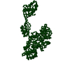
|
|---|---|
| 1 |
|
-要素
| #1: タンパク質 | 分子量: 93407.125 Da / 分子数: 1 / 由来タイプ: 天然 / 由来: (天然) |
|---|
-実験情報
-実験
| 実験 | 手法: 電子顕微鏡法 |
|---|---|
| EM実験 | 試料の集合状態: PARTICLE / 3次元再構成法: 単粒子再構成法 |
- 試料調製
試料調製
| 構成要素 | 名称: eEF2-bound 80S complex in presence of AlF4-, and GDP タイプ: RIBOSOME |
|---|---|
| 緩衝液 | 名称: 20 mM Hepes-NH3, 100 mM KCl, 20 mM MgCl2 / pH: 7.2 / 詳細: 20 mM Hepes-NH3, 100 mM KCl, 20 mM MgCl2 |
| 試料 | 包埋: NO / シャドウイング: NO / 染色: NO / 凍結: YES |
| 急速凍結 | 装置: FEI VITROBOT MARK I / 凍結剤: ETHANE |
- 電子顕微鏡撮影
| 実験機器 |  モデル: Tecnai F20 / 画像提供: FEI Company |
|---|---|
| 顕微鏡 | モデル: FEI TECNAI F20 |
| 電子銃 | 電子線源:  FIELD EMISSION GUN / 加速電圧: 200 kV / 照射モード: FLOOD BEAM FIELD EMISSION GUN / 加速電圧: 200 kV / 照射モード: FLOOD BEAM |
| 電子レンズ | モード: BRIGHT FIELD / 倍率(公称値): 50000 X / 倍率(補正後): 49650 X / 最大 デフォーカス(公称値): 1500 nm / 最小 デフォーカス(公称値): 4500 nm |
| 試料ホルダ | 温度: 93 K |
| 撮影 | 電子線照射量: 10 e/Å2 / フィルム・検出器のモデル: KODAK SO-163 FILM |
- 解析
解析
| EMソフトウェア |
| ||||||||||||
|---|---|---|---|---|---|---|---|---|---|---|---|---|---|
| CTF補正 | 詳細: segregation in defocus groups and correction in volumes | ||||||||||||
| 対称性 | 点対称性: C1 (非対称) | ||||||||||||
| 3次元再構成 | 手法: Single particle 3D reconstruction / 解像度: 12.6 Å / ピクセルサイズ(公称値): 2.82 Å 詳細: supervised classification was used (ref. Valle, M. et al, 2002, EMBO J.) 対称性のタイプ: POINT | ||||||||||||
| 原子モデル構築 | プロトコル: RIGID BODY FIT / 空間: REAL 詳細: METHOD--Each domain fitted as rigid body, domains I (G and G), II,IV, and V were fitted. Domain III, and domain IV insertions were not included in this fitting model. The coordinates for this ...詳細: METHOD--Each domain fitted as rigid body, domains I (G and G), II,IV, and V were fitted. Domain III, and domain IV insertions were not included in this fitting model. The coordinates for this entry are based on manual fitting of the coordinates of the crystal structure 1N0U into cryo-EM density map. Therefore, authors did not deposit new structure factors. | ||||||||||||
| 原子モデル構築 | PDB-ID: 1N0U Accession code: 1N0U / Source name: PDB / タイプ: experimental model | ||||||||||||
| 精密化ステップ | サイクル: LAST
|
