ムービー
ムービー コントローラー


+ データを開く

- 基本情報
基本情報
| 登録情報 | データベース: EMDB / ID: EMD-8364 | |||||||||
|---|---|---|---|---|---|---|---|---|---|---|
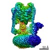
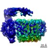
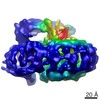
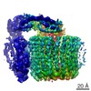
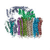
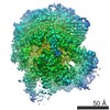
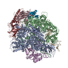
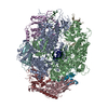
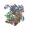
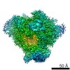
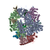
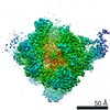
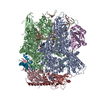
| タイトル | CryoEM map of the S. cerevisiae Vo region solubilized in DDM | |||||||||
 マップデータ マップデータ | Map of the Yeast Vo region solubilized in DDM | |||||||||
 試料 試料 |
| |||||||||
| 機能・相同性 |  機能・相同性情報 機能・相同性情報cell wall mannoprotein biosynthetic process / ATPase-coupled ion transmembrane transporter activity / cellular response to alkaline pH / P-type proton-exporting transporter activity / Insulin receptor recycling / Transferrin endocytosis and recycling / polyphosphate metabolic process / ROS and RNS production in phagocytes / Amino acids regulate mTORC1 / Golgi lumen acidification ...cell wall mannoprotein biosynthetic process / ATPase-coupled ion transmembrane transporter activity / cellular response to alkaline pH / P-type proton-exporting transporter activity / Insulin receptor recycling / Transferrin endocytosis and recycling / polyphosphate metabolic process / ROS and RNS production in phagocytes / Amino acids regulate mTORC1 / Golgi lumen acidification / vacuolar transport / vacuolar proton-transporting V-type ATPase, V0 domain / endosomal lumen acidification / protein targeting to vacuole / proton-transporting V-type ATPase complex / vacuolar proton-transporting V-type ATPase complex / vacuole organization / fungal-type vacuole / vacuolar acidification / cellular hyperosmotic response / fungal-type vacuole membrane / phosphatidylinositol-3,5-bisphosphate binding / proton transmembrane transporter activity / intracellular copper ion homeostasis / Neutrophil degranulation / proton-transporting ATPase activity, rotational mechanism / proton transmembrane transport / cell periphery / transmembrane transport /  エンドサイトーシス / ATPase binding / protein-containing complex assembly / intracellular iron ion homeostasis / ゴルジ体 / 生体膜 エンドサイトーシス / ATPase binding / protein-containing complex assembly / intracellular iron ion homeostasis / ゴルジ体 / 生体膜類似検索 - 分子機能 | |||||||||
| 生物種 |  Saccharomyces cerevisiae (パン酵母) Saccharomyces cerevisiae (パン酵母) | |||||||||
| 手法 | 単粒子再構成法 / クライオ電子顕微鏡法 / 解像度: 8.3 Å | |||||||||
 データ登録者 データ登録者 | Mazhab-Jafari MT / Rohou A / Schmidt C / Bueler SA / Benlekbir S / Robinson C / Rubinstein JL | |||||||||
 引用 引用 | ジャーナル: Nature / 年: 2016 タイトル: Atomic model for the membrane-embedded V motor of a eukaryotic V-ATPase. 著者: Mohammad T Mazhab-Jafari / Alexis Rohou / Carla Schmidt / Stephanie A Bueler / Samir Benlekbir / Carol V Robinson / John L Rubinstein /    要旨: Vacuolar-type ATPases (V-ATPases) are ATP-powered proton pumps involved in processes such as endocytosis, lysosomal degradation, secondary transport, TOR signalling, and osteoclast and kidney ...Vacuolar-type ATPases (V-ATPases) are ATP-powered proton pumps involved in processes such as endocytosis, lysosomal degradation, secondary transport, TOR signalling, and osteoclast and kidney function. ATP hydrolysis in the soluble catalytic V region drives proton translocation through the membrane-embedded V region via rotation of a rotor subcomplex. Variability in the structure of the intact enzyme has prevented construction of an atomic model for the membrane-embedded motor of any rotary ATPase. We induced dissociation and auto-inhibition of the V and V regions of the V-ATPase by starving the yeast Saccharomyces cerevisiae, allowing us to obtain a ~3.9-Å resolution electron cryomicroscopy map of the V complex and build atomic models for the majority of its subunits. The analysis reveals the structures of subunits acc'c″de and a protein that we identify and propose to be a new subunit (subunit f). A large cavity between subunit a and the c-ring creates a cytoplasmic half-channel for protons. The c-ring has an asymmetric distribution of proton-carrying Glu residues, with the Glu residue of subunit c″ interacting with Arg735 of subunit a. The structure suggests sequential protonation and deprotonation of the c-ring, with ATP-hydrolysis-driven rotation causing protonation of a Glu residue at the cytoplasmic half-channel and subsequent deprotonation of a Glu residue at a luminal half-channel. | |||||||||
| 履歴 |
|
- 構造の表示
構造の表示
| ムービー |
ムービービューア |
|---|---|
| 構造ビューア | EMマップ: SurfViewMolmilJmol/JSmol |
| 添付画像 |

- ダウンロードとリンク
-EMDBアーカイブ
| マップデータ | emd_8364.map.gz | 14.5 MB | EMDBマップデータ形式 | |
|---|---|---|---|---|
| ヘッダ (付随情報) | emd-8364-v30.xmlemd-8364.xml | 10.3 KB 10.3 KB | 表示 表示 | EMDBヘッダ |
| FSC (解像度算出) | emd_8364_fsc.xml | 5.6 KB | 表示 | FSCデータファイル |
| 画像 | 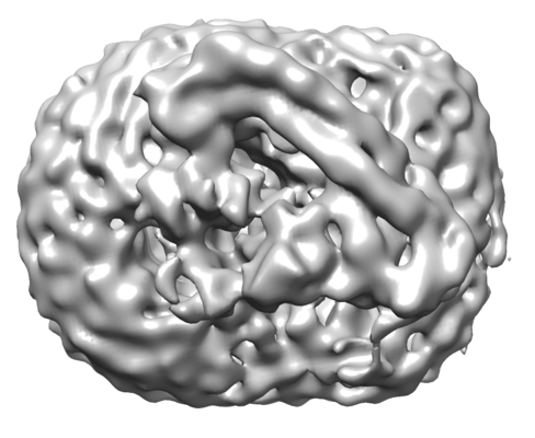 emd_8364.png emd_8364.png | 95.7 KB | ||
| アーカイブディレクトリ |  http://ftp.pdbj.org/pub/emdb/structures/EMD-8364ftp://ftp.pdbj.org/pub/emdb/structures/EMD-8364 http://ftp.pdbj.org/pub/emdb/structures/EMD-8364ftp://ftp.pdbj.org/pub/emdb/structures/EMD-8364 | HTTPS FTP |
-関連構造データ
-リンク
| EMDBのページ | EMDB (EBI/PDBe) / EMDataResource |
|---|---|
| 「今月の分子」の関連する項目 |


-マップ
| ファイル | ダウンロード / ファイル: emd_8364.map.gz / 形式: CCP4 / 大きさ: 15.6 MB / タイプ: IMAGE STORED AS FLOATING POINT NUMBER (4 BYTES) | ||||||||||||||||||||||||||||||||||||||||||||||||||||||||||||||||||||
|---|---|---|---|---|---|---|---|---|---|---|---|---|---|---|---|---|---|---|---|---|---|---|---|---|---|---|---|---|---|---|---|---|---|---|---|---|---|---|---|---|---|---|---|---|---|---|---|---|---|---|---|---|---|---|---|---|---|---|---|---|---|---|---|---|---|---|---|---|---|
| 注釈 | Map of the Yeast Vo region solubilized in DDM | ||||||||||||||||||||||||||||||||||||||||||||||||||||||||||||||||||||
| ボクセルのサイズ | X=Y=Z: 1.45 Å | ||||||||||||||||||||||||||||||||||||||||||||||||||||||||||||||||||||
| 密度 |
| ||||||||||||||||||||||||||||||||||||||||||||||||||||||||||||||||||||
| 対称性 | 空間群: 1 | ||||||||||||||||||||||||||||||||||||||||||||||||||||||||||||||||||||
| 詳細 | EMDB XML:
CCP4マップ ヘッダ情報:
| ||||||||||||||||||||||||||||||||||||||||||||||||||||||||||||||||||||
-添付データ
- 試料の構成要素
-全体 : Vo region
| 全体 | 名称: Vo region |
|---|---|
| 要素 |
|
-超分子 #1: Vo region
| 超分子 | 名称: Vo region / タイプ: complex / ID: 1 / 親要素: 0 / 含まれる分子: #1 詳細: Vo region of Saccharomyces cerevisiae Vacuolar-type ATPase solubilized in DDM |
|---|---|
| 由来(天然) | 生物種: Saccharomyces cerevisiae (パン酵母) |
| 分子量 | 理論値: 300 KDa |
-実験情報
-構造解析
| 手法 | クライオ電子顕微鏡法 |
|---|---|
 解析 解析 | 単粒子再構成法 |
| 試料の集合状態 | particle |
-試料調製
| 濃度 | 2.5 mg/mL |
|---|---|
| 緩衝液 | pH: 7.5 |
| 凍結 | 凍結剤: ETHANE-PROPANE / チャンバー内湿度: 100 % / 装置: FEI VITROBOT MARK III |
| 詳細 | Vo solubilized in DDM Buffer: 50 mM Tris-HCl pH 7.5, 150 mM NaCl, 0.02 % [w/v] DDM |
- 電子顕微鏡法
| 顕微鏡 | FEI TECNAI F20 |
|---|---|
| 電子線 | 加速電圧: 200 kV / 電子線源: FIELD EMISSION GUN |
| 電子光学系 | 照射モード: FLOOD BEAM / 撮影モード: BRIGHT FIELDBright-field microscopy |
| 試料ステージ | 試料ホルダーモデル: GATAN LIQUID NITROGEN |
| 撮影 | フィルム・検出器のモデル: GATAN K2 SUMMIT (4k x 4k) 検出モード: COUNTING / デジタル化 - 画像ごとのフレーム数: 1-30 / 撮影したグリッド数: 6 / 平均露光時間: 15.0 sec. / 平均電子線量: 36.0 e/Å2 |
| 実験機器 |  モデル: Tecnai F20 / 画像提供: FEI Company |
-画像解析
| CTF補正 | ソフトウェア - 名称: CTFFIND3 |
|---|---|
| 初期モデル | モデルのタイプ: EMDB MAP EMDB ID: 詳細: The Vo region was segmented out and used as the initial model |
| 初期 角度割当 | タイプ: OTHER / ソフトウェア - 名称: RELION (ver. 1.3) |
| 最終 角度割当 | タイプ: OTHER / ソフトウェア - 名称: RELION (ver. 1.3) |
| 最終 再構成 | 想定した対称性 - 点群: C1 (非対称) / 解像度のタイプ: BY AUTHOR / 解像度: 8.3 Å / 解像度の算出法: FSC 0.143 CUT-OFF / ソフトウェア - 名称: RELION (ver. 1.3) / 使用した粒子像数: 43184 |
| FSC曲線 (解像度の算出) | 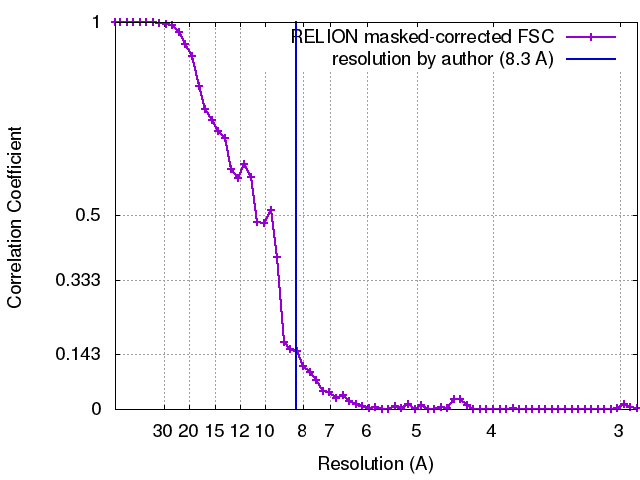 |