Movie
Movie Controller
 Structure viewers
 About Yorodumi Papers


+Search query
-Structure paper
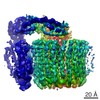
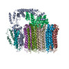
| Title | Atomic model for the membrane-embedded V motor of a eukaryotic V-ATPase. |
|---|---|
| Journal, issue, pages | Nature, Vol. 539, Issue 7627, Page 118-122, Year 2016 |
| Publish date | Nov 3, 2016 |
 Authors Authors | Mohammad T Mazhab-Jafari / Alexis Rohou / Carla Schmidt / Stephanie A Bueler / Samir Benlekbir / Carol V Robinson / John L Rubinstein /    |
| PubMed Abstract | Vacuolar-type ATPases (V-ATPases) are ATP-powered proton pumps involved in processes such as endocytosis, lysosomal degradation, secondary transport, TOR signalling, and osteoclast and kidney ...Vacuolar-type ATPases (V-ATPases) are ATP-powered proton pumps involved in processes such as endocytosis, lysosomal degradation, secondary transport, TOR signalling, and osteoclast and kidney function. ATP hydrolysis in the soluble catalytic V region drives proton translocation through the membrane-embedded V region via rotation of a rotor subcomplex. Variability in the structure of the intact enzyme has prevented construction of an atomic model for the membrane-embedded motor of any rotary ATPase. We induced dissociation and auto-inhibition of the V and V regions of the V-ATPase by starving the yeast Saccharomyces cerevisiae, allowing us to obtain a ~3.9-Å resolution electron cryomicroscopy map of the V complex and build atomic models for the majority of its subunits. The analysis reveals the structures of subunits acc'c″de and a protein that we identify and propose to be a new subunit (subunit f). A large cavity between subunit a and the c-ring creates a cytoplasmic half-channel for protons. The c-ring has an asymmetric distribution of proton-carrying Glu residues, with the Glu residue of subunit c″ interacting with Arg735 of subunit a. The structure suggests sequential protonation and deprotonation of the c-ring, with ATP-hydrolysis-driven rotation causing protonation of a Glu residue at the cytoplasmic half-channel and subsequent deprotonation of a Glu residue at a luminal half-channel. |
 External links External links | Nature / PubMed:27776355 / PubMed Central |
| Methods | EM (single particle) |
| Resolution | 3.9 - 8.7 Å |
| Structure data | 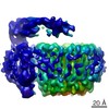 EMDB-8363: 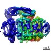 EMDB-8364: 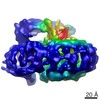 EMDB-8367: |
| Source |
|
 Keywords Keywords |  MOTOR PROTEIN / Rotary ATPase / Vacuolar-type ATPase / Electron Cryomicroscopy / Vo region / Membrane protein MOTOR PROTEIN / Rotary ATPase / Vacuolar-type ATPase / Electron Cryomicroscopy / Vo region / Membrane protein |