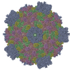
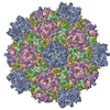
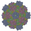
ジャーナル: Structure / 年: 2011 タイトル: Atomic model of CPV reveals the mechanism used by this single-shelled virus to economically carry out functions conserved in multishelled reoviruses. 著者: Xuekui Yu / Peng Ge / Jiansen Jiang / Ivo Atanasov / Z Hong Zhou / 要旨: Unlike the multishelled viruses in the Reoviridae, cytoplasmic polyhedrosis virus (CPV) is single shelled, yet stable and fully capable of carrying out functions conserved within Reoviridae. Here, we ...Unlike the multishelled viruses in the Reoviridae, cytoplasmic polyhedrosis virus (CPV) is single shelled, yet stable and fully capable of carrying out functions conserved within Reoviridae. Here, we report a 3.1 Å resolution cryo electron microscopy structure of CPV and derive its atomic model, consisting of 60 turret proteins (TPs), 120 each of capsid shell proteins (CSPs) and large protrusion proteins (LPPs). Two unique segments of CSP contribute to CPV's stability: an inserted protrusion domain interacting with neighboring proteins, and an N-anchor tying up CSPs together through strong interactions such as β sheet augmentation. Without the need to interact with outer shell proteins, LPP retains only the N-terminal two-third region containing a conserved helix-barrel core and interacts exclusively with CSP. TP is also simplified, containing only domains involved in RNA capping. Our results illustrate how CPV proteins have evolved in a coordinative manner to economically carry out their conserved functions.
 ムービー
ムービー コントローラー


 データを開く
 基本情報
基本情報 マップデータ
 試料
試料 キーワード
キーワード cytoplasmic polyhedrosis virus /
  機能・相同性情報

 データ登録者
 引用
引用
 構造の表示
構造の表示
 ダウンロードとリンク
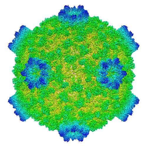 emd_5256_1.jpg
 http://ftp.pdbj.org/pub/emdb/structures/EMD-5256

 試料の構成要素
 解析
解析 電子顕微鏡法
