 Movie
Movie Controller


+ Open data

- Basic information
| Entry | Database: PDB / ID: 3izx | ||||||
|---|---|---|---|---|---|---|---|
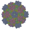
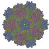
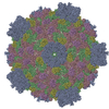
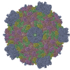
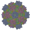
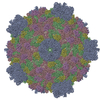
| Title | 3.1 Angstrom cryoEM structure of cytoplasmic polyhedrosis virus | ||||||
 Components Components |
| ||||||
 Keywords Keywords |  VIRUS / cytoplasmic polyhedrosis virus / 3D reconstruction / cryoEM / full atom model VIRUS / cytoplasmic polyhedrosis virus / 3D reconstruction / cryoEM / full atom model | ||||||
| Function / homology |  Function and homology information Function and homology information | ||||||
| Biological species |   Bombyx mori cypovirus 1 Bombyx mori cypovirus 1 | ||||||
| Method | ELECTRON MICROSCOPY / single particle reconstruction / cryo EM / Resolution: 3.1 Å | ||||||
 Authors Authors | Yu, X. / Ge, P. / Jiang, J. / Atanasov, I. / Zhou, Z.H. | ||||||
 Citation Citation | Journal: Structure / Year: 2011 Title: Atomic model of CPV reveals the mechanism used by this single-shelled virus to economically carry out functions conserved in multishelled reoviruses. Authors: Xuekui Yu / Peng Ge / Jiansen Jiang / Ivo Atanasov / Z Hong Zhou /  Abstract: Unlike the multishelled viruses in the Reoviridae, cytoplasmic polyhedrosis virus (CPV) is single shelled, yet stable and fully capable of carrying out functions conserved within Reoviridae. Here, we ...Unlike the multishelled viruses in the Reoviridae, cytoplasmic polyhedrosis virus (CPV) is single shelled, yet stable and fully capable of carrying out functions conserved within Reoviridae. Here, we report a 3.1 Å resolution cryo electron microscopy structure of CPV and derive its atomic model, consisting of 60 turret proteins (TPs), 120 each of capsid shell proteins (CSPs) and large protrusion proteins (LPPs). Two unique segments of CSP contribute to CPV's stability: an inserted protrusion domain interacting with neighboring proteins, and an N-anchor tying up CSPs together through strong interactions such as β sheet augmentation. Without the need to interact with outer shell proteins, LPP retains only the N-terminal two-third region containing a conserved helix-barrel core and interacts exclusively with CSP. TP is also simplified, containing only domains involved in RNA capping. Our results illustrate how CPV proteins have evolved in a coordinative manner to economically carry out their conserved functions. | ||||||
| History |
|
- Structure visualization
| Movie |
Movie viewer |
|---|---|
| Structure viewer | Molecule: MolmilJmol/JSmol |
- Downloads & links
-Download
| PDBx/mmCIF format | 3izx.cif.gz | 744.1 KB | Display | PDBx/mmCIF format |
|---|---|---|---|---|
| PDB format | pdb3izx.ent.gz | 612.4 KB | Display | PDB format |
| PDBx/mmJSON format | 3izx.json.gz | Tree view | PDBx/mmJSON format | |
| Others |  Other downloads Other downloads |
-Validation report
| Arichive directory | https://data.pdbj.org/pub/pdb/validation_reports/iz/3izxftp://data.pdbj.org/pub/pdb/validation_reports/iz/3izx | HTTPS FTP |
|---|
-Related structure data
| Related structure data | 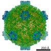 5256MC M: map data used to model this data C: citing same article ( |
|---|---|
| Similar structure data |
-Links
 PDBj
PDBj
- Assembly
| Deposited unit | 
|
|---|---|
| 1 | x 60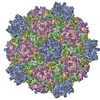
|
| 2 |
|
| 3 | x 5
|
| 4 | x 6
|
| 5 | 
|
| Symmetry | Point symmetry: (Schoenflies symbol: I (icosahedral)) |
-Components
| #1: Protein | Structure Mass: 120145.797 Da / Num. of mol.: 1 / Source method: isolated from a natural source / Source: (natural) Bombyx mori cypovirus 1 / References: UniProt: Q914N6 | ||
|---|---|---|---|
| #2: Protein | Mass: 148560.859 Da / Num. of mol.: 2 / Source method: isolated from a natural source / Source: (natural) Bombyx mori cypovirus 1 / References: UniProt: Q6TS43#3: Protein | Mass: 49929.211 Da / Num. of mol.: 2 / Source method: isolated from a natural source / Source: (natural) Bombyx mori cypovirus 1 / References: UniProt: C6K2M8 |
-Experimental details
-Experiment
| Experiment | Method: ELECTRON MICROSCOPY |
|---|---|
| EM experiment | Aggregation state: PARTICLE / 3D reconstruction method: single particle reconstruction |
- Sample preparation
| Component | Name: cytoplasmic polyhedrosis virus (CPV) / Type: VIRUS Details: Icosahedral particle. CPV particles were purified from polyhedra. |
|---|---|
| Details of virus | Empty: NO / Enveloped: NO / Host category: INVERTERBRATES / Isolate: STRAIN / Type: VIRION |
| Buffer solution | pH: 7.4 / Details: 10mM PBS |
| Specimen | Conc.: 3 mg/ml / Embedding applied: NO / Shadowing applied: NO / Staining applied: NO / Vitrification applied: YES / Details: 10mM PBS |
| Vitrification | Cryogen name: ETHANE / Temp: 90 K / Humidity: 40 % / Method: Blot for 3 seconds before plunging |
- Electron microscopy imaging
| Experimental equipment |  Model: Titan Krios / Image courtesy: FEI Company |
|---|---|
| Microscopy | Model: FEI TITAN KRIOS / Date: Jul 12, 2010 / Details: Low dose |
| Electron gun | Electron source: FIELD EMISSION GUN / Accelerating voltage: 300 kV / Illumination mode: FLOOD BEAM / Electron beam tilt params: 0 |
| Electron lens | Mode: BRIGHT FIELDBright-field microscopy / Nominal magnification: 59000 X / Nominal defocus max: 2500 nm / Nominal defocus min: 1000 nm / Cs: 2.75 mm Astigmatism : objective lens astigmatism was corrected a t 150,000 magnificationCamera length: 0 mm |
| Specimen holder | Specimen holder model: OTHER / Specimen holder type: Eucentric / Temperature: 90 K / Tilt angle max: -9999 ° / Tilt angle min: -9999 ° |
| Image recording | Electron dose: 25 e/Å2 / Film or detector model: KODAK SO-163 FILM |
- Processing
| EM software | Name: IMIRS / Category: 3D reconstruction | ||||||||||||
|---|---|---|---|---|---|---|---|---|---|---|---|---|---|
| CTF correction | Details: Each particle | ||||||||||||
| Symmetry | Point symmetry: I (icosahedral) | ||||||||||||
| 3D reconstruction | Method: common line / Resolution: 3.1 Å / Resolution method: FSC 0.143 CUT-OFF / Num. of particles: 28993 Details: ( Details about the particle: 645 micrographs that clearly showed signals beyond 6 angstrom in their spectra ) Symmetry type: POINT | ||||||||||||
| Refinement step | Cycle: LAST
|
