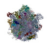
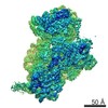
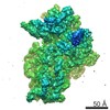
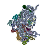
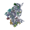
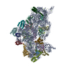
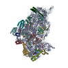
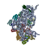
ジャーナル: Nucleic Acids Res / 年: 2017 タイトル: Cryo-EM structure of the spinach chloroplast ribosome reveals the location of plastid-specific ribosomal proteins and extensions. 著者: Michael Graf / Stefan Arenz / Paul Huter / Alexandra Dönhöfer / Jirí Novácek / Daniel N Wilson / 要旨: Ribosomes are the protein synthesizing machines of the cell. Recent advances in cryo-EM have led to the determination of structures from a variety of species, including bacterial 70S and eukaryotic ...Ribosomes are the protein synthesizing machines of the cell. Recent advances in cryo-EM have led to the determination of structures from a variety of species, including bacterial 70S and eukaryotic 80S ribosomes as well as mitoribosomes from eukaryotic mitochondria, however, to date high resolution structures of plastid 70S ribosomes have been lacking. Here we present a cryo-EM structure of the spinach chloroplast 70S ribosome, with an average resolution of 5.4 Å for the small 30S subunit and 3.6 Å for the large 50S ribosomal subunit. The structure reveals the location of the plastid-specific ribosomal proteins (RPs) PSRP1, PSRP4, PSRP5 and PSRP6 as well as the numerous plastid-specific extensions of the RPs. We discover many features by which the plastid-specific extensions stabilize the ribosome via establishing additional interactions with surrounding ribosomal RNA and RPs. Moreover, we identify a large conglomerate of plastid-specific protein mass adjacent to the tunnel exit site that could facilitate interaction of the chloroplast ribosome with the thylakoid membrane and the protein-targeting machinery. Comparing the Escherichia coli 70S ribosome with that of the spinach chloroplast ribosome provides detailed insight into the co-evolution of RP and rRNA.
 ムービー
ムービー コントローラー


 データを開く
 基本情報
基本情報 マップデータ
 試料
試料
 Spinacia oleracea (ホウレンソウ)
 データ登録者
 引用
引用

 構造の表示
構造の表示 ムービービューア
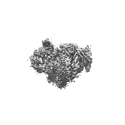
 ダウンロードとリンク
 emd_3526.png
 http://ftp.pdbj.org/pub/emdb/structures/EMD-3526
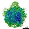


 試料の構成要素
 解析
解析 電子顕微鏡法