 ムービー
ムービー コントローラー


+ データを開く

- 基本情報
基本情報
| 登録情報 | データベース: EMDB / ID: EMD-2380 | |||||||||
|---|---|---|---|---|---|---|---|---|---|---|
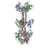
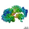
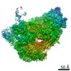
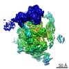
| タイトル | Structure of herpesvirus fusion glycoprotein B-bilayer complex revealing the protein-membrane and lateral protein-protein interaction | |||||||||
 マップデータ マップデータ | Subtomogram average of HSV-1 glycoprotein B bound to a lipid bilayer | |||||||||
 試料 試料 |
| |||||||||
 キーワード キーワード | electron cryo microscopy /  tomography (トモグラフィー) / membrane proximal region / protein coat / pseudo-atomic / modelling / virus-host interaction tomography (トモグラフィー) / membrane proximal region / protein coat / pseudo-atomic / modelling / virus-host interaction | |||||||||
| 機能・相同性 |  機能・相同性情報 機能・相同性情報host cell Golgi membrane / host cell endosome membrane / symbiont entry into host cell / エンベロープ (ウイルス) / virion attachment to host cell / host cell plasma membrane / virion membrane / 生体膜 / identical protein binding類似検索 - 分子機能 | |||||||||
| 生物種 |   Human herpesvirus 1 (ヘルペスウイルス) Human herpesvirus 1 (ヘルペスウイルス) | |||||||||
| 手法 | サブトモグラム平均法 / クライオ電子顕微鏡法 / 解像度: 27.0 Å | |||||||||
 データ登録者 データ登録者 | Maurer UE / Zeev-Ben-Mordehai Z / Pandurangan AP / Cairns TM / Hannah BP / Whitbeck JC / Eisenberg RJ / Cohen GH / Topf M / Huiskonen JT / Grunewald K | |||||||||
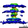
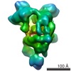
 引用 引用 | ジャーナル: Structure / 年: 2013 タイトル: The structure of herpesvirus fusion glycoprotein B-bilayer complex reveals the protein-membrane and lateral protein-protein interaction. 著者: Ulrike E Maurer / Tzviya Zeev-Ben-Mordehai / Arun Prasad Pandurangan / Tina M Cairns / Brian P Hannah / J Charles Whitbeck / Roselyn J Eisenberg / Gary H Cohen / Maya Topf / Juha T Huiskonen / Kay Grünewald /  要旨: Glycoprotein B (gB) is a key component of the complex herpesvirus fusion machinery. We studied membrane interaction of two gB ectodomain forms and present an electron cryotomography structure of the ...Glycoprotein B (gB) is a key component of the complex herpesvirus fusion machinery. We studied membrane interaction of two gB ectodomain forms and present an electron cryotomography structure of the gB-bilayer complex. The two forms differed in presence or absence of the membrane proximal region (MPR) but showed an overall similar trimeric shape. The presence of the MPR impeded interaction with liposomes. In contrast, the MPR-lacking form interacted efficiently with liposomes. Lateral interaction resulted in coat formation on the membranes. The structure revealed that interaction of gB with membranes was mediated by the fusion loops and limited to the outer membrane leaflet. The observed intrinsic propensity of gB to cluster on membranes indicates an additional role of gB in driving the fusion process forward beyond the transient fusion pore opening and subsequently leading to fusion pore expansion. | |||||||||
| 履歴 |
|
- 構造の表示
構造の表示
| ムービー |
ムービービューア |
|---|---|
| 構造ビューア | EMマップ: SurfViewMolmilJmol/JSmol |
| 添付画像 |

- ダウンロードとリンク
-EMDBアーカイブ
| マップデータ | emd_2380.map.gz | 3.5 MB | EMDBマップデータ形式 | |
|---|---|---|---|---|
| ヘッダ (付随情報) | emd-2380-v30.xmlemd-2380.xml | 11.8 KB 11.8 KB | 表示 表示 | EMDBヘッダ |
| 画像 | 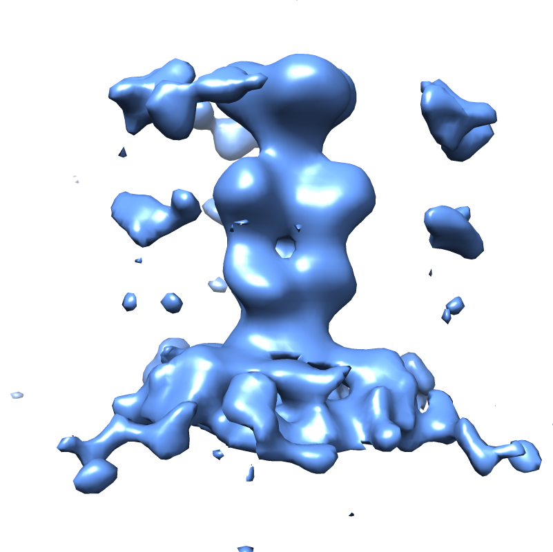 EMD-2380.png EMD-2380.png | 228.7 KB | ||
| アーカイブディレクトリ |  http://ftp.pdbj.org/pub/emdb/structures/EMD-2380ftp://ftp.pdbj.org/pub/emdb/structures/EMD-2380 http://ftp.pdbj.org/pub/emdb/structures/EMD-2380ftp://ftp.pdbj.org/pub/emdb/structures/EMD-2380 | HTTPS FTP |
-関連構造データ
-リンク
| EMDBのページ | EMDB (EBI/PDBe) / EMDataResource |
|---|
-マップ
| ファイル | ダウンロード / ファイル: emd_2380.map.gz / 形式: CCP4 / 大きさ: 3.7 MB / タイプ: IMAGE STORED AS FLOATING POINT NUMBER (4 BYTES) | ||||||||||||||||||||||||||||||||||||||||||||||||||||||||||||||||||||
|---|---|---|---|---|---|---|---|---|---|---|---|---|---|---|---|---|---|---|---|---|---|---|---|---|---|---|---|---|---|---|---|---|---|---|---|---|---|---|---|---|---|---|---|---|---|---|---|---|---|---|---|---|---|---|---|---|---|---|---|---|---|---|---|---|---|---|---|---|---|
| 注釈 | Subtomogram average of HSV-1 glycoprotein B bound to a lipid bilayer | ||||||||||||||||||||||||||||||||||||||||||||||||||||||||||||||||||||
| ボクセルのサイズ | X=Y=Z: 4.6 Å | ||||||||||||||||||||||||||||||||||||||||||||||||||||||||||||||||||||
| 密度 |
| ||||||||||||||||||||||||||||||||||||||||||||||||||||||||||||||||||||
| 対称性 | 空間群: 1 | ||||||||||||||||||||||||||||||||||||||||||||||||||||||||||||||||||||
| 詳細 | EMDB XML:
CCP4マップ ヘッダ情報:
| ||||||||||||||||||||||||||||||||||||||||||||||||||||||||||||||||||||
-添付データ
- 試料の構成要素
-全体 : HSV-1 glycoprotein B ectodomain lacking the membrane-proximal reg...
| 全体 | 名称: HSV-1 glycoprotein B ectodomain lacking the membrane-proximal region bound to a lipid bilayer |
|---|---|
| 要素 |
|
-超分子 #1000: HSV-1 glycoprotein B ectodomain lacking the membrane-proximal reg...
| 超分子 | 名称: HSV-1 glycoprotein B ectodomain lacking the membrane-proximal region bound to a lipid bilayer タイプ: sample / ID: 1000 / 集合状態: One trimer of gB bound to a lipid bilayer / Number unique components: 2 |
|---|---|
| 分子量 | 実験値: 259 KDa / 理論値: 235 KDa 手法: Weights are for the trimeric ectodomain lacking the membrane proximal region. The experimental weight was determined by mass spectrometry. The theoretical weight was calculated from the sequence. |
-分子 #1: Envelope glycoprotein B
| 分子 | 名称: Envelope glycoprotein B / タイプ: protein_or_peptide / ID: 1 / Name.synonym: gB-1, gB1 詳細: Liposomes consisting of phosphatidylcholine and cholesterol at 1.7:1 molar ratio were incubated with gB at pH 5.5 at 37C for one hour 集合状態: Trimer / 組換発現: Yes |
|---|---|
| 由来(天然) | 生物種: Human herpesvirus 1 (ヘルペスウイルス) / 株: stain KOS / 別称: Human herpes simplex virus 1 |
| 組換発現 | 生物種:   Spodoptera frugiperda (ツマジロクサヨトウ) Spodoptera frugiperda (ツマジロクサヨトウ)組換細胞: Sf9 / 組換プラスミド: pCW289 |
| 配列 | UniProtKB: Envelope glycoprotein B / GO: symbiont entry into host cell / InterPro: Herpesvirus Glycoprotein B |
-実験情報
-構造解析
| 手法 | クライオ電子顕微鏡法 |
|---|---|
 解析 解析 | サブトモグラム平均法 |
| 試料の集合状態 | particle |
-試料調製
| 濃度 | 1.0 mg/mL |
|---|---|
| 緩衝液 | pH: 5.5 / 詳細: PBS with sodium citrate |
| グリッド | 詳細: Cflat |
| 凍結 | 凍結剤: ETHANE-PROPANE MIXTURE / チャンバー内温度: 120 K / 装置: OTHER / 手法: Blot manually for 3 s before plunging |
- 電子顕微鏡法
| 顕微鏡 | FEI TECNAI F20 |
|---|---|
| 電子線 | 加速電圧: 200 kV / 電子線源: FIELD EMISSION GUN |
| 電子光学系 | 倍率(補正後): 67000 / 照射モード: FLOOD BEAM / 撮影モード: BRIGHT FIELDBright-field microscopy / Cs: 2 mm / 最大 デフォーカス(公称値): 2.0 µm / 最小 デフォーカス(公称値): 2.0 µm |
| 試料ステージ | 試料ホルダー: liquid nitrogen cooled / 試料ホルダーモデル: SIDE ENTRY, EUCENTRIC / Tilt series - Axis1 - Min angle: -60 ° / Tilt series - Axis1 - Max angle: 60 ° |
| 日付 | 2008年1月17日 |
| 撮影 | カテゴリ: CCD フィルム・検出器のモデル: GATAN ULTRASCAN 4000 (4k x 4k) デジタル化 - サンプリング間隔: 15 µm / 実像数: 9 / 平均電子線量: 100 e/Å2 詳細: The dataset consists of 9 tomograms (containing 38 liposomes with bound gB). Data were binned by factor of 2. ビット/ピクセル: 12 |
| 実験機器 |  モデル: Tecnai F20 / 画像提供: FEI Company |
-画像解析
| CTF補正 | 詳細: Low pass filter to the first zero crossing of the CTF |
|---|---|
| 最終 再構成 | 想定した対称性 - 点群: C3 (3回回転対称) / アルゴリズム: OTHER / 解像度のタイプ: BY AUTHOR / 解像度: 27.0 Å / 解像度の算出法: FSC 0.5 CUT-OFF / ソフトウェア - 名称: Jsubtomo 詳細: The best 786 spikes (of 996) were selected based on constrained cross correlation coefficient and by excluding overlaps. All three Euler angles of the spike were refined. 使用したサブトモグラム数: 996 |
| 詳細 | The sub-tomograms were picked manually from tomographic reconstructions of 38 liposomes |

