- EMDB-2023: Three-dimensional reconstruction of a targeted individual 17nm na... -
+
データを開く
IDまたはキーワード:
読み込み中...
-
基本情報
登録情報
データベース: EMDB / ID: EMD-2023
タイトル
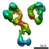
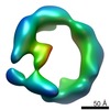
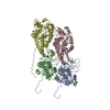
Three-dimensional reconstruction of a targeted individual 17nm nascent high-density lipoprotein (HDL) particle by individual-particle electron tomography (IPET). The reconstruction displayed a ring-shaped structure of containing three protein - apoipoprotein A-Is (28 kDa each).
ジャーナル: PLoS One / 年: 2012 タイトル: IPET and FETR: experimental approach for studying molecular structure dynamics by cryo-electron tomography of a single-molecule structure. 著者: Lei Zhang / Gang Ren / 要旨: The dynamic personalities and structural heterogeneity of proteins are essential for proper functioning. Structural determination of dynamic/heterogeneous proteins is limited by conventional ...The dynamic personalities and structural heterogeneity of proteins are essential for proper functioning. Structural determination of dynamic/heterogeneous proteins is limited by conventional approaches of X-ray and electron microscopy (EM) of single-particle reconstruction that require an average from thousands to millions different molecules. Cryo-electron tomography (cryoET) is an approach to determine three-dimensional (3D) reconstruction of a single and unique biological object such as bacteria and cells, by imaging the object from a series of tilting angles. However, cconventional reconstruction methods use large-size whole-micrographs that are limited by reconstruction resolution (lower than 20 Å), especially for small and low-symmetric molecule (<400 kDa). In this study, we demonstrated the adverse effects from image distortion and the measuring tilt-errors (including tilt-axis and tilt-angle errors) both play a major role in limiting the reconstruction resolution. Therefore, we developed a "focused electron tomography reconstruction" (FETR) algorithm to improve the resolution by decreasing the reconstructing image size so that it contains only a single-instance protein. FETR can tolerate certain levels of image-distortion and measuring tilt-errors, and can also precisely determine the translational parameters via an iterative refinement process that contains a series of automatically generated dynamic filters and masks. To describe this method, a set of simulated cryoET images was employed; to validate this approach, the real experimental images from negative-staining and cryoET were used. Since this approach can obtain the structure of a single-instance molecule/particle, we named it individual-particle electron tomography (IPET) as a new robust strategy/approach that does not require a pre-given initial model, class averaging of multiple molecules or an extended ordered lattice, but can tolerate small tilt-errors for high-resolution single "snapshot" molecule structure determination. Thus, FETR/IPET provides a completely new opportunity for a single-molecule structure determination, and could be used to study the dynamic character and equilibrium fluctuation of macromolecules.
詳細: Tomography tilt angle from -60 to 60 in step of 1.5
最終 再構成
アルゴリズム: OTHER / 解像度のタイプ: BY AUTHOR / 解像度: 42.0 Å / 解像度の算出法: OTHER / ソフトウェア - 名称: IPET, and, FETR 詳細: Map was reconstructed by individual-particle electron tomography (IPET)and Focus ET Reconstruction Algorithm.
詳細
Single targeted particle"s images were reconstructed by focus ET reconstruction (FETR) algorithm of individual particle electron tomography (IPET) method. Average number of tilts used in the 3D reconstructions: 81. Average tomographic tilt angle increment: 1.5.
 ムービー
ムービー コントローラー


 データを開く
 基本情報
基本情報 マップデータ
 試料
試料 キーワード
キーワード
 Homo sapiens (ヒト)
 データ登録者
 引用
引用
 構造の表示
構造の表示 ムービービューア
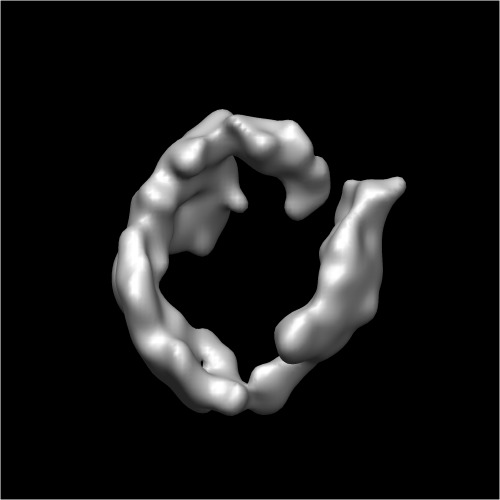
 ダウンロードとリンク
 http://ftp.pdbj.org/pub/emdb/structures/EMD-2023
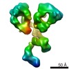
 試料の構成要素
 解析
解析 電子顕微鏡法