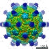
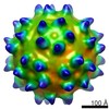
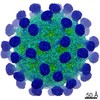
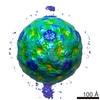
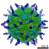
登録情報 データベース : EMDB / ID : EMD-1114タイトル The crystal structure of coxsackievirus A21 and its interaction with ICAM-1. This is an cryo-EM reconstructed map of human coxsackievirus A21 complex with 5 domain ICAM-1kilifi 試料 : Human Coxsackievirus A21 complex with ICAM-1KilifiFcウイルス : タンパク質・ペプチド : ICAM-1KilifiFc機能・相同性 分子機能 ドメイン・相同性 構成要素
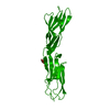
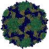
/ / / / / / / / / / / / / / / / / / / / / / / / / / / / / / / / / / / / / / / / / / / / / / / / / / / / / / / / / / / / / / / / / / / / / / / / / / / / / / / / / / / / / / / / / / / / / / / / / / / / / / / / / / / / / / / / / / / / / / / / / / / / / / / / / / / 生物種 手法 / / 解像度 : 8.0 Å Xiao C / Bator-Kelly CM / Rieder E / Chipman PR / Craig A / Kuhn RJ / Wimmer E / Rossmann MG ジャーナル : Structure / 年 : 2005タイトル : The crystal structure of coxsackievirus A21 and its interaction with ICAM-1.著者 : Chuan Xiao / Carol M Bator-Kelly / Elizabeth Rieder / Paul R Chipman / Alister Craig / Richard J Kuhn / Eckard Wimmer / Michael G Rossmann / 要旨 : CVA21 and polioviruses both belong to the Enterovirus genus in the family of Picornaviridae, whereas rhinoviruses form a distinct picornavirus genus. Nevertheless, CVA21 and the major group of human ... CVA21 and polioviruses both belong to the Enterovirus genus in the family of Picornaviridae, whereas rhinoviruses form a distinct picornavirus genus. Nevertheless, CVA21 and the major group of human rhinoviruses recognize intercellular adhesion molecule-1 (ICAM-1) as their cellular receptor, whereas polioviruses use poliovirus receptor. The crystal structure of CVA21 has been determined to 3.2 A resolution. Its structure has greater similarity to poliovirus structures than to other known picornavirus structures. Cryo-electron microscopy (cryo-EM) was used to determine an 8.0 A resolution structure of CVA21 complexed with an ICAM-1 variant, ICAM-1(Kilifi). The cryo-EM map was fitted with the crystal structures of ICAM-1 and CVA21. Significant differences in the structure of CVA21 with respect to the poliovirus structures account for the inability of ICAM-1 to bind polioviruses. The interface between CVA21 and ICAM-1 has shape and electrostatic complementarity with many residues being conserved among those CVAs that bind ICAM-1. 履歴 登録 2005年3月29日 - ヘッダ(付随情報) 公開 2005年3月29日 - マップ公開 2005年10月4日 - 更新 2011年5月26日 - 現状 2011年5月26日 処理サイト : PDBe / 状態 : 公開
すべて表示 表示を減らす
 ムービー
ムービー コントローラー


 データを開く
 基本情報
基本情報 マップデータ
 試料
試料 機能・相同性情報
 免疫シナプス / Integrin cell surface interactions / negative regulation of endothelial cell apoptotic process / negative regulation of extrinsic apoptotic signaling pathway via death domain receptors /
 データ登録者
 引用
引用
 構造の表示
構造の表示
 ダウンロードとリンク
 1114.gif
 http://ftp.pdbj.org/pub/emdb/structures/EMD-1114
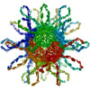


 試料の構成要素
 解析
解析 電子顕微鏡法