regulation of translational termination / translation release factor activity, codon nonspecific / translation release factor activity, codon specific / guanosine tetraphosphate binding / stringent response / mRNA base-pairing translational repressor activity / ornithine decarboxylase inhibitor activity / transcription antitermination factor activity, RNA binding / misfolded RNA binding / Group I intron splicing ...regulation of translational termination / translation release factor activity, codon nonspecific / translation release factor activity, codon specific / guanosine tetraphosphate binding / stringent response / mRNA base-pairing translational repressor activity / ornithine decarboxylase inhibitor activity / transcription antitermination factor activity, RNA binding / misfolded RNA binding / Group I intron splicing / RNA folding / transcriptional attenuation / endoribonuclease inhibitor activity / RNA-binding transcription regulator activity / positive regulation of ribosome biogenesis / negative regulation of cytoplasmic translation / translational termination / DnaA-L2 complex / four-way junction DNA binding / translation repressor activity / negative regulation of translational initiation / negative regulation of DNA-templated DNA replication initiation / regulation of mRNA stability / ribosome assembly / mRNA regulatory element binding translation repressor activity / assembly of large subunit precursor of preribosome / positive regulation of RNA splicing / transcription elongation factor complex / response to reactive oxygen species / DNA endonuclease activity / cytosolic ribosome assembly / regulation of DNA-templated transcription elongation / transcription antitermination / regulation of cell growth / maintenance of translational fidelity / DNA-templated transcription termination / response to radiation / mRNA 5'-UTR binding / ribosomal small subunit biogenesis / ribosomal large subunit assembly / small ribosomal subunit rRNA binding / ribosomal small subunit assembly / GDP binding / cytosolic small ribosomal subunit / large ribosomal subunit rRNA binding / ribosome binding / large ribosomal subunit / リボソーム生合成 / regulation of translation / small ribosomal subunit / cytoplasmic translation / 5S rRNA binding / cytosolic large ribosomal subunit / transferase activity / tRNA binding / negative regulation of translation / rRNA binding / molecular adaptor activity / リボソーム / structural constituent of ribosome / 翻訳 (生物学) / response to antibiotic / mRNA binding / GTPase activity / negative regulation of DNA-templated transcription / GTP binding / DNA binding / RNA binding / zinc ion binding / 生体膜 / 細胞質基質 / 細胞質 類似検索 - 分子機能
Apidaecin / Apidaecin / Peptide chain release factor 3, C-terminal / Peptide chain release factor 3, domain III superfamily / Peptide chain release factor 3, GTP-binding domain / Class II release factor RF3, C-terminal domain / Peptide chain release factor 3 / Peptide chain release factor 1 / Peptide chain release factor / PCRF domain ...Apidaecin / Apidaecin / Peptide chain release factor 3, C-terminal / Peptide chain release factor 3, domain III superfamily / Peptide chain release factor 3, GTP-binding domain / Class II release factor RF3, C-terminal domain / Peptide chain release factor 3 / Peptide chain release factor 1 / Peptide chain release factor / PCRF domain / PCRF / Peptide chain release factor class I superfamily / Prokaryotic-type class I peptide chain release factors signature. / Peptide chain release factor class I / RF-1 domain / Ribosomal protein L10, eubacterial, conserved site / Ribosomal protein L10 signature. / Ribosomal protein L10 / EF-G domain III/V-like / : / Tr-type G domain, conserved site / Translational (tr)-type guanine nucleotide-binding (G) domain signature. / Translation elongation factor EFTu-like, domain 2 / Ribosomal protein S21, conserved site / Ribosomal protein S21 signature. / Ribosomal protein L25, short-form / Ribosomal protein S14, bacterial/plastid / Elongation factor Tu domain 2 / Ribosomal protein L11, bacterial-type / Ribosomal protein S21 superfamily / Ribosomal protein S21 / Ribosomal protein S16, conserved site / Ribosomal protein S16 signature. / Translational (tr)-type GTP-binding domain / Elongation factor Tu GTP binding domain / Translational (tr)-type guanine nucleotide-binding (G) domain profile. / Ribosomal protein S21 / Ribosomal protein L11, conserved site / Ribosomal protein L10-like domain superfamily / Ribosomal protein L21, conserved site / Ribosomal protein L21 signature. / Ribosomal protein L11 signature. / Ribosomal protein L10P / Ribosomal protein L10 / Ribosomal protein L16 signature 1. / : / Ribosomal protein L6, conserved site / Ribosomal protein L6 signature 1. / Ribosomal protein L16, conserved site / Ribosomal protein L16 signature 2. / Ribosomal protein L11, N-terminal / Ribosomal protein L9 signature. / Ribosomal protein L17 signature. / Ribosomal protein L9, bacteria/chloroplast / Ribosomal protein L9, C-terminal / Ribosomal protein L9, C-terminal domain / Ribosomal protein L9, C-terminal domain superfamily / Ribosomal protein L11/L12 / Ribosomal protein L11, C-terminal / Ribosomal protein L11, C-terminal domain superfamily / Ribosomal protein L11/L12, N-terminal domain superfamily / Ribosomal protein L11/L12 / Ribosomal protein L11, N-terminal domain / Ribosomal protein L11, RNA binding domain / Ribosomal L25p family / Ribosomal protein L25 / Ribosomal protein L28/L24 superfamily / Ribosomal protein L36 signature. / Ribosomal protein L25/Gln-tRNA synthetase, N-terminal / Ribosomal protein L32p, bacterial type / Ribosomal protein L25/Gln-tRNA synthetase, anti-codon-binding domain superfamily / Ribosomal protein L9, N-terminal domain superfamily / Ribosomal protein L9 / Ribosomal protein L9, N-terminal / Ribosomal protein L9, N-terminal domain / Ribosomal protein L28 / Ribosomal protein L35, conserved site / Ribosomal protein L35 signature. / Ribosomal protein L33, conserved site / Ribosomal protein L33 signature. / Ribosomal protein L35, non-mitochondrial / Ribosomal protein L5, bacterial-type / Ribosomal protein L6, bacterial-type / Ribosomal protein L18, bacterial-type / Ribosomal protein L19, conserved site / Ribosomal protein L19 signature. / Ribosomal protein L36 / Ribosomal protein L36 superfamily / Ribosomal protein L36 / Ribosomal protein L9/RNase H1, N-terminal / Ribosomal protein L20 signature. / Ribosomal protein S3, bacterial-type / Ribosomal protein S6, conserved site / Ribosomal protein S6 signature. / Ribosomal protein L27, conserved site / Ribosomal protein L27 signature. / Ribosomal protein S19, bacterial-type / Ribosomal protein S7, bacterial/organellar-type / Ribosomal protein S11, bacterial-type / Ribosomal protein S13, bacterial-type 類似検索 - ドメイン・相同性
Small ribosomal subunit protein bS6 / Small ribosomal subunit protein uS7 / Large ribosomal subunit protein uL15 / Peptide chain release factor RF1 / Peptide chain release factor RF3 / Large ribosomal subunit protein uL10 / Large ribosomal subunit protein uL11 / Large ribosomal subunit protein bL19 / Large ribosomal subunit protein bL20 / Large ribosomal subunit protein bL27 ...Small ribosomal subunit protein bS6 / Small ribosomal subunit protein uS7 / Large ribosomal subunit protein uL15 / Peptide chain release factor RF1 / Peptide chain release factor RF3 / Large ribosomal subunit protein uL10 / Large ribosomal subunit protein uL11 / Large ribosomal subunit protein bL19 / Large ribosomal subunit protein bL20 / Large ribosomal subunit protein bL27 / Large ribosomal subunit protein bL28 / Large ribosomal subunit protein uL29 / Large ribosomal subunit protein bL32 / Large ribosomal subunit protein bL33 / Large ribosomal subunit protein bL34 / Large ribosomal subunit protein bL35 / Large ribosomal subunit protein bL36A / Large ribosomal subunit protein bL9 / Small ribosomal subunit protein uS10 / Small ribosomal subunit protein uS11 / Small ribosomal subunit protein uS12 / Small ribosomal subunit protein uS13 / Small ribosomal subunit protein bS16 / Small ribosomal subunit protein bS18 / Small ribosomal subunit protein uS19 / Small ribosomal subunit protein bS20 / Small ribosomal subunit protein uS2 / Small ribosomal subunit protein uS3 / Small ribosomal subunit protein uS4 / Small ribosomal subunit protein uS5 / Small ribosomal subunit protein uS8 / Small ribosomal subunit protein uS9 / Large ribosomal subunit protein uL13 / Large ribosomal subunit protein uL14 / Large ribosomal subunit protein uL16 / Large ribosomal subunit protein uL23 / Small ribosomal subunit protein uS15 / Large ribosomal subunit protein bL17 / Large ribosomal subunit protein bL21 / Large ribosomal subunit protein uL30 / Large ribosomal subunit protein uL6 / Small ribosomal subunit protein uS14 / Small ribosomal subunit protein uS17 / Large ribosomal subunit protein uL18 / Large ribosomal subunit protein uL2 / Large ribosomal subunit protein uL3 / Large ribosomal subunit protein uL24 / Large ribosomal subunit protein uL4 / Large ribosomal subunit protein uL22 / Large ribosomal subunit protein uL5 / Small ribosomal subunit protein bS21 / Large ribosomal subunit protein bL25 / Apidaecin 類似検索 - 構成要素
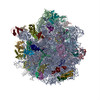
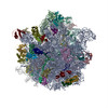
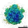
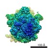
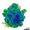
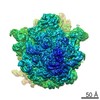
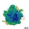
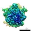
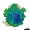
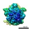
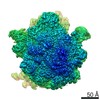
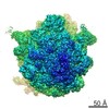
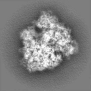
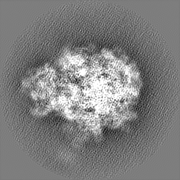
ジャーナル: Nat Commun / 年: 2018 タイトル: Visualization of translation termination intermediates trapped by the Apidaecin 137 peptide during RF3-mediated recycling of RF1. 著者: Michael Graf / Paul Huter / Cristina Maracci / Miroslav Peterek / Marina V Rodnina / Daniel N Wilson / 要旨: During translation termination in bacteria, the release factors RF1 and RF2 are recycled from the ribosome by RF3. While high-resolution structures of the individual termination factors on the ...During translation termination in bacteria, the release factors RF1 and RF2 are recycled from the ribosome by RF3. While high-resolution structures of the individual termination factors on the ribosome exist, direct structural insight into how RF3 mediates dissociation of the decoding RFs has been lacking. Here we have used the Apidaecin 137 peptide to trap RF1 together with RF3 on the ribosome and visualize an ensemble of termination intermediates using cryo-electron microscopy. Binding of RF3 to the ribosome induces small subunit (SSU) rotation and swivelling of the head, yielding intermediate states with shifted P-site tRNAs and RF1 conformations. RF3 does not directly eject RF1 from the ribosome, but rather induces full rotation of the SSU that indirectly dislodges RF1 from its binding site. SSU rotation is coupled to the accommodation of the GTPase domain of RF3 on the large subunit (LSU), thereby promoting GTP hydrolysis and dissociation of RF3 from the ribosome.
 ムービー
ムービー コントローラー


 データを開く
 基本情報
基本情報 マップデータ
 試料
試料 キーワード
キーワード GTPase (GTPアーゼ) /
  機能・相同性情報

 データ登録者
 ドイツ,
ドイツ,  チェコ, 4件
  引用
引用 構造の表示
構造の表示
 ダウンロードとリンク
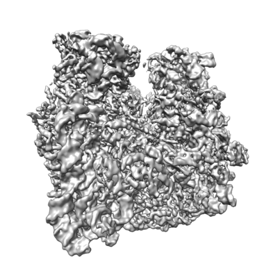 emd_0081.png
 http://ftp.pdbj.org/pub/emdb/structures/EMD-0081


 Z
Z Y
Y X
X


 試料の構成要素
 解析
解析 電子顕微鏡法