 ムービー
ムービー コントローラー


+ データを開く

- 基本情報
基本情報
| 登録情報 | データベース: EMDB / ID: EMD-2563 | |||||||||
|---|---|---|---|---|---|---|---|---|---|---|
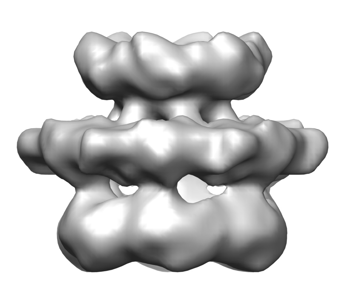
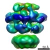
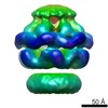
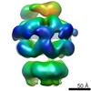
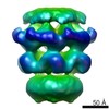
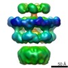
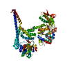
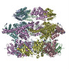
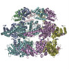
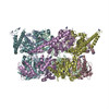
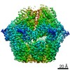
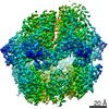
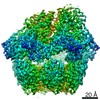
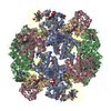
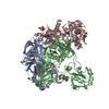
| タイトル | Cryo electron microscopy of E. coli ClpB | |||||||||
 マップデータ マップデータ | Cryo-EM reconstruction of E.coli ClpB. Six fold symmetry applied. | |||||||||
 試料 試料 |
| |||||||||
 キーワード キーワード |  chaperone (シャペロン) / disaggregase / ClpB / coiled-coil domain (コイルドコイル) chaperone (シャペロン) / disaggregase / ClpB / coiled-coil domain (コイルドコイル) | |||||||||
| 生物種 |  Escherichia coli (大腸菌) Escherichia coli (大腸菌) | |||||||||
| 手法 | 単粒子再構成法 / クライオ電子顕微鏡法 / 解像度: 29.0 Å | |||||||||
 データ登録者 データ登録者 | Carroni M / Kummer E / Oguchi Y / Clare DK / Wendler P / Sinning I / Kopp J / Mogk A / Bukau B / Saibil HR | |||||||||
 引用 引用 | ジャーナル: Elife / 年: 2014 タイトル: Head-to-tail interactions of the coiled-coil domains regulate ClpB activity and cooperation with Hsp70 in protein disaggregation. 著者: Marta Carroni / Eva Kummer / Yuki Oguchi / Petra Wendler / Daniel K Clare / Irmgard Sinning / Jürgen Kopp / Axel Mogk / Bernd Bukau / Helen R Saibil /   要旨: The hexameric AAA+ chaperone ClpB reactivates aggregated proteins in cooperation with the Hsp70 system. Essential for disaggregation, the ClpB middle domain (MD) is a coiled-coil propeller that binds ...The hexameric AAA+ chaperone ClpB reactivates aggregated proteins in cooperation with the Hsp70 system. Essential for disaggregation, the ClpB middle domain (MD) is a coiled-coil propeller that binds Hsp70. Although the ClpB subunit structure is known, positioning of the MD in the hexamer and its mechanism of action are unclear. We obtained electron microscopy (EM) structures of the BAP variant of ClpB that binds the protease ClpP, clearly revealing MD density on the surface of the ClpB ring. Mutant analysis and asymmetric reconstructions show that MDs adopt diverse positions in a single ClpB hexamer. Adjacent, horizontally oriented MDs form head-to-tail contacts and repress ClpB activity by preventing Hsp70 interaction. Tilting of the MD breaks this contact, allowing Hsp70 binding, and releasing the contact in adjacent subunits. Our data suggest a wavelike activation of ClpB subunits around the ring.DOI: http://dx.doi.org/10.7554/eLife.02481.001. | |||||||||
| 履歴 |
|
- 構造の表示
構造の表示
| ムービー |
 ムービービューア ムービービューア |
|---|---|
| 構造ビューア | EMマップ: SurfViewMolmilJmol/JSmol |
| 添付画像 |
- ダウンロードとリンク
-EMDBアーカイブ
| マップデータ | emd_2563.map.gz | 7.2 MB | EMDBマップデータ形式 | |
|---|---|---|---|---|
| ヘッダ (付随情報) | emd-2563-v30.xmlemd-2563.xml | 11.1 KB 11.1 KB | 表示 表示 | EMDBヘッダ |
| FSC (解像度算出) | emd_2563_fsc.xml | 5.4 KB | 表示 | FSCデータファイル |
| 画像 | emd_2563.tiff | 328.2 KB | ||
| アーカイブディレクトリ |  http://ftp.pdbj.org/pub/emdb/structures/EMD-2563ftp://ftp.pdbj.org/pub/emdb/structures/EMD-2563 http://ftp.pdbj.org/pub/emdb/structures/EMD-2563ftp://ftp.pdbj.org/pub/emdb/structures/EMD-2563 | HTTPS FTP |
-関連構造データ
-リンク
| EMDBのページ | EMDB (EBI/PDBe) / EMDataResource |
|---|
-マップ
| ファイル | ダウンロード / ファイル: emd_2563.map.gz / 形式: CCP4 / 大きさ: 7.8 MB / タイプ: IMAGE STORED AS FLOATING POINT NUMBER (4 BYTES) | ||||||||||||||||||||||||||||||||||||||||||||||||||||||||||||||||||||
|---|---|---|---|---|---|---|---|---|---|---|---|---|---|---|---|---|---|---|---|---|---|---|---|---|---|---|---|---|---|---|---|---|---|---|---|---|---|---|---|---|---|---|---|---|---|---|---|---|---|---|---|---|---|---|---|---|---|---|---|---|---|---|---|---|---|---|---|---|---|
| 注釈 | Cryo-EM reconstruction of E.coli ClpB. Six fold symmetry applied. | ||||||||||||||||||||||||||||||||||||||||||||||||||||||||||||||||||||
| ボクセルのサイズ | X=Y=Z: 2 Å | ||||||||||||||||||||||||||||||||||||||||||||||||||||||||||||||||||||
| 密度 |
| ||||||||||||||||||||||||||||||||||||||||||||||||||||||||||||||||||||
| 対称性 | 空間群: 1 | ||||||||||||||||||||||||||||||||||||||||||||||||||||||||||||||||||||
| 詳細 | EMDB XML:
CCP4マップ ヘッダ情報:
| ||||||||||||||||||||||||||||||||||||||||||||||||||||||||||||||||||||
-添付データ
- 試料の構成要素
-全体 : ClpB with ATPgammaS.
| 全体 | 名称: ClpB with ATPgammaS. |
|---|---|
| 要素 |
|
-超分子 #1000: ClpB with ATPgammaS.
| 超分子 | 名称: ClpB with ATPgammaS. / タイプ: sample / ID: 1000 / 集合状態: Homohexamer. / Number unique components: 1 |
|---|---|
| 分子量 | 理論値: 500 KDa |
-分子 #1: ClpB
| 分子 | 名称: ClpB / タイプ: protein_or_peptide / ID: 1 / コピー数: 6 / 集合状態: Hexamer / 組換発現: Yes |
|---|---|
| 由来(天然) | 生物種: Escherichia coli (大腸菌) / 細胞中の位置: cytoplasm |
| 分子量 | 実験値: 80 KDa / 理論値: 80 KDa |
| 組換発現 | 生物種: Escherichia coli (大腸菌) / 組換株: derivatives of MC4100 / 組換プラスミド: pDS56 |
-実験情報
-構造解析
| 手法 | クライオ電子顕微鏡法 |
|---|---|
 解析 解析 | 単粒子再構成法 |
| 試料の集合状態 | particle |
-試料調製
| 濃度 | 0.3 mg/mL |
|---|---|
| 緩衝液 | pH: 7.5 詳細: 20 mM Tris-HCl, pH 7.5, 20 mM KCl, 15 mM MgCl2, 1 mM DTT, 2 mM ATPgammaS |
| 凍結 | 凍結剤: ETHANE / チャンバー内湿度: 100 % / チャンバー内温度: 77 K / 装置: FEI VITROBOT MARK II |
- 電子顕微鏡法
| 顕微鏡 | FEI TECNAI F20 |
|---|---|
| 電子線 | 加速電圧: 200 kV / 電子線源: FIELD EMISSION GUN |
| 電子光学系 | 照射モード: FLOOD BEAM / 撮影モード: BRIGHT FIELDBright-field microscopy / Cs: 2 mm / 最大 デフォーカス(公称値): 4.0 µm / 最小 デフォーカス(公称値): 1.5 µm / 倍率(公称値): 80000 |
| 試料ステージ | 試料ホルダーモデル: SIDE ENTRY, EUCENTRIC |
| 温度 | 最低: 77 K / 最高: 88 K / 平均: 83 K |
| アライメント法 | Legacy - 非点収差: Objective lens astigmatism was corrected at 150,000 x magnification |
| 日付 | 2011年2月4日 |
| 撮影 | カテゴリ: CCD フィルム・検出器のモデル: GATAN ULTRASCAN 4000 (4k x 4k) 実像数: 200 / 平均電子線量: 15 e/Å2 |
| 実験機器 |  モデル: Tecnai F20 / 画像提供: FEI Company |
-画像解析
| CTF補正 | 詳細: phase flipping entire frame |
|---|---|
| 最終 角度割当 | 詳細: Only side views used selected by using MSA and classification. Beta angles between 80 and 100 degrees (IMAGIC convention). |
| 最終 再構成 | 想定した対称性 - 点群: C6 (6回回転対称) / アルゴリズム: OTHER / 解像度のタイプ: BY AUTHOR / 解像度: 29.0 Å / 解像度の算出法: OTHER / ソフトウェア - 名称: IMAGIC, Spider 詳細: Starting models were generated by angular reconstitution and particle orientations were refined by projection matching in SPIDER. 使用した粒子像数: 7606 |
| FSC曲線 (解像度の算出) | 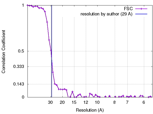 |
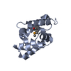
-原子モデル構築 1
| 初期モデル | PDB ID: Chain - Chain ID: A |
|---|---|
| ソフトウェア | 名称: Chimera |
| 詳細 | Fitting of separate domains was performed manually and locally optimised using Chimera. Known domain interfaces were used to guide the fit. |
| 精密化 | 空間: REAL / プロトコル: RIGID BODY FIT |
-原子モデル構築 2
| 初期モデル | PDB ID: Chain - Chain ID: A |
|---|---|
| ソフトウェア | 名称: Chimera |
| 詳細 | Fitting of separate domains was performed manually and locally optimised using Chimera. Known domain interfaces were used to guide the fit. |
| 精密化 | 空間: REAL / プロトコル: RIGID BODY FIT |
