The French Muscular Dystrophy Telethon (AFM-Telethon)
23207
フランス
The French Muscular Dystrophy Telethon (AFM-Telethon)
23210
フランス
引用
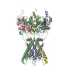
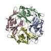
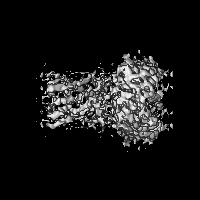
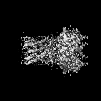
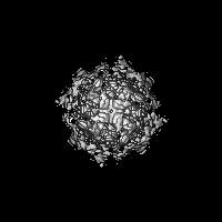
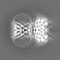
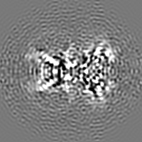
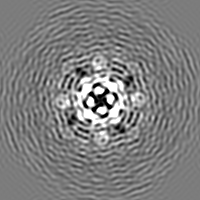
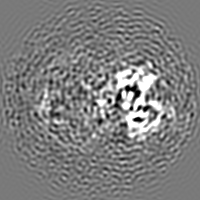
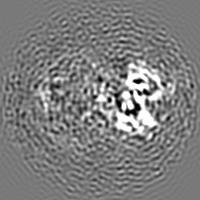
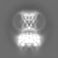
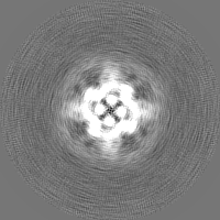
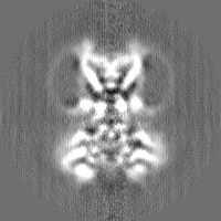
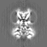
ジャーナル: Sci Adv / 年: 2022 タイトル: Cryo-electron microscopy unveils unique structural features of the human Kir2.1 channel. 著者: Carlos A H Fernandes / Dania Zuniga / Charline Fagnen / Valérie Kugler / Rosa Scala / Gérard Péhau-Arnaudet / Renaud Wagner / David Perahia / Saïd Bendahhou / Catherine Vénien-Bryan / 要旨: We present the first structure of the human Kir2.1 channel containing both transmembrane domain (TMD) and cytoplasmic domain (CTD). Kir2.1 channels are strongly inward-rectifying potassium channels ...We present the first structure of the human Kir2.1 channel containing both transmembrane domain (TMD) and cytoplasmic domain (CTD). Kir2.1 channels are strongly inward-rectifying potassium channels that play a key role in maintaining resting membrane potential. Their gating is modulated by phosphatidylinositol 4,5-bisphosphate (PIP). Genetically inherited defects in Kir2.1 channels are responsible for several rare human diseases, including Andersen's syndrome. The structural analysis (cryo-electron microscopy), surface plasmon resonance, and electrophysiological experiments revealed a well-connected network of interactions between the PIP-binding site and the G-loop through residues R312 and H221. In addition, molecular dynamics simulations and normal mode analysis showed the intrinsic tendency of the CTD to tether to the TMD and a movement of the secondary anionic binding site to the membrane even without PIP. Our results revealed structural features unique to human Kir2.1 and provided insights into the connection between G-loop and gating and the pathological mechanisms associated with this channel.
For structural fitting, it was used dock-in-map (available at PHENIX) that uses both SSM and convolution-based shape searches to find a part of a map that is similar to a model. An initial in silico homology model of human Kir2.1 was generated using I-TASSER using the crystal structure of chicken Kir2.2 channel (PDB ID 3JYC) as a template. For building and refinement of the atomic model, the transmembrane domain (TMD, 55-184 region) of this in silico model was placed into the final sharpened cryo-EM map using the Dock in Map tool available in PHENIX. For the cytoplasmic domain (CTD; 188-367 region), the crystal structure of the CTD from mice Kir2.1 channel (PDB ID 1U4F) was placed into the final cryo-EM map using the same approach. Once the models were placed in the electron density, the loops that connect the two domains (185-187 region) and a N-terminal loop (41-54 region) absent in the in silico model were manually built using Coot.
精密化
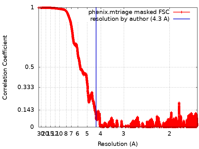
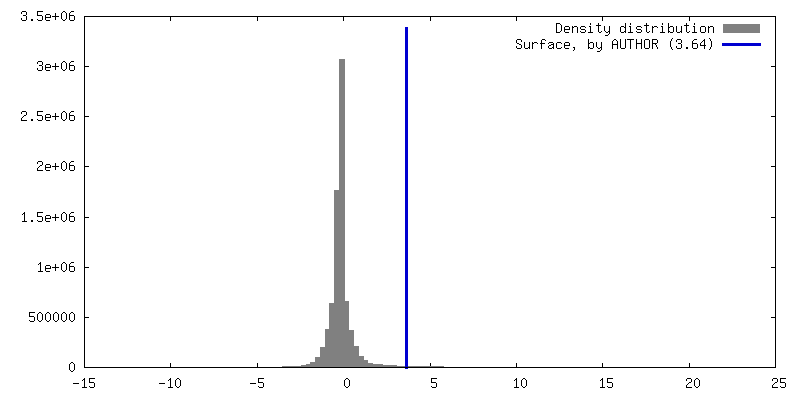
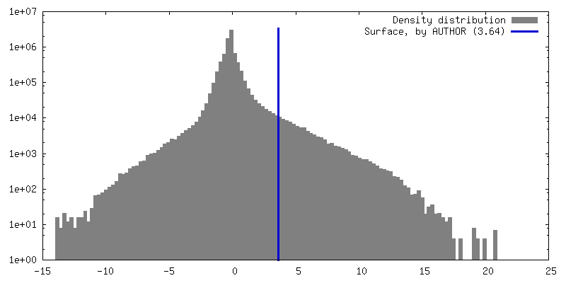
空間: REAL / プロトコル: OTHER / 温度因子: 404.73 / 当てはまり具合の基準: Correlation coefficient
得られたモデル
PDB-7zdz: Cryo-EM structure of the human inward-rectifier potassium 2.1 channel (Kir2.1)
 ムービー
ムービー コントローラー


 データを開く
 基本情報
基本情報
 マップデータ
 試料
試料 機能・相同性情報
 regulation of resting membrane potential ...Sensory perception of sour taste / Classical Kir channels / regulation of skeletal muscle contraction via regulation of action potential / relaxation of skeletal muscle / Phase 4 - resting membrane potential / magnesium ion transport / voltage-gated potassium channel activity involved in cardiac muscle cell action potential repolarization / membrane repolarization during action potential / membrane repolarization during cardiac muscle cell action potential /
 データ登録者
 フランス, 3件
  引用
引用 構造の表示
構造の表示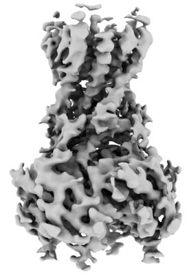
 ダウンロードとリンク
 emd_14678.png
 http://ftp.pdbj.org/pub/emdb/structures/EMD-14678
 X (Sec.)
 Y (Row.)
 Z (Col.)


 試料の構成要素

 解析
解析 電子顕微鏡法