 Movie
Movie Controller

+ Open data

- Basic information
| Entry | Database: SASBDB / ID: SASDA32 |
|---|---|
 Sample Sample | BSA in HEPES
|
| Function / homology |  Function and homology information Function and homology informationcellular response to calcium ion starvation /  enterobactin binding / negative regulation of mitochondrial depolarization / toxic substance binding / small molecule binding / cellular response to starvation / fatty acid binding / pyridoxal phosphate binding / blood microparticle / protein-containing complex ...cellular response to calcium ion starvation / enterobactin binding / negative regulation of mitochondrial depolarization / toxic substance binding / small molecule binding / cellular response to starvation / fatty acid binding / pyridoxal phosphate binding / blood microparticle / protein-containing complex / DNA binding / extracellular region / metal ion binding / cytoplasm enterobactin binding / negative regulation of mitochondrial depolarization / toxic substance binding / small molecule binding / cellular response to starvation / fatty acid binding / pyridoxal phosphate binding / blood microparticle / protein-containing complex ...cellular response to calcium ion starvation / enterobactin binding / negative regulation of mitochondrial depolarization / toxic substance binding / small molecule binding / cellular response to starvation / fatty acid binding / pyridoxal phosphate binding / blood microparticle / protein-containing complex / DNA binding / extracellular region / metal ion binding / cytoplasmSimilarity search - Function |
| Biological species |  Bos taurus (cattle) Bos taurus (cattle) |
 Contact author Contact author |
|
- Structure visualization
| Structure viewer | Molecule: MolmilJmol/JSmol |
|---|
- Downloads & links
-Data source
| SASBDB page |  SASDA32 SASDA32 |
|---|
-Related structure data
| Similar structure data |
|---|


-External links
| Related items in Molecule of the Month |
|---|


-Models
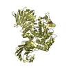
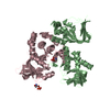
| Model #4 |   Type: dummy / Radius of dummy atoms: 1.80 A / Symmetry : P1 / Chi-square value: 2.566404 Search similar-shape structures of this assembly by Omokage search (details) Search similar-shape structures of this assembly by Omokage search (details) |
|---|---|
| Model #3 |  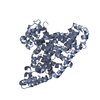 Type: atomic / Software: (28) Search similar-shape structures of this assembly by Omokage search (details) |
-Sample
| Sample | Name: BSA in HEPES / Ext coefficient: 6.46 / Contrast: 2.953 / Specific vol: 0.737 / Purity method: gel filtration / Dry vol: 17570 / Sample MW: 66 kDa / Specimen concentration: 25.64 mg/ml / Concentration method: nanodrop |
|---|---|
| Buffer | Name: 50 mM HEPES 50 mM KCl / Concentration: 50.00 mM / PK: 7 / pH: 7.5 / Comment: 4-(2-hydroxyethyl)-1-piperazineethanesulfonic acid / Composition: KCl 50.000 mM |
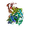
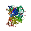
| Entity #1 | Name: BSA / Type: protein / Description: Serum albumin / Formula weight: 69.29 / Num. of mol.: 1 / Source: Bos taurus / References: UniProt: P02769 Sequence: MKWVTFISLL LLFSSAYSRG VFRRDTHKSE IAHRFKDLGE EHFKGLVLIA FSQYLQQCPF DEHVKLVNEL TEFAKTCVAD ESHAGCEKSL HTLFGDELCK VASLRETYGD MADCCEKQEP ERNECFLSHK DDSPDLPKLK PDPNTLCDEF KADEKKFWGK YLYEIARRHP ...Sequence: MKWVTFISLL LLFSSAYSRG VFRRDTHKSE IAHRFKDLGE EHFKGLVLIA FSQYLQQCPF DEHVKLVNEL TEFAKTCVAD ESHAGCEKSL HTLFGDELCK VASLRETYGD MADCCEKQEP ERNECFLSHK DDSPDLPKLK PDPNTLCDEF KADEKKFWGK YLYEIARRHP YFYAPELLYY ANKYNGVFQE CCQAEDKGAC LLPKIETMRE KVLASSARQR LRCASIQKFG ERALKAWSVA RLSQKFPKAE FVEVTKLVTD LTKVHKECCH GDLLECADDR ADLAKYICDN QDTISSKLKE CCDKPLLEKS HCIAEVEKDA IPENLPPLTA DFAEDKDVCK NYQEAKDAFL GSFLYEYSRR HPEYAVSVLL RLAKEYEATL EECCAKDDPH ACYSTVFDKL KHLVDEPQNL IKQNCDQFEK LGEYGFQNAL IVRYTRKVPQ VSTPTLVEVS RSLGKVGTRC CTKPESERMP CTEDYLSLIL NRLCVLHEKT PVSEKVTKCC TESLVNRRPC FSALTPDETY VPKAFDEKLF TFHADICTLP DTEKQIKKQT ALVELLKHKP KATEEQLKTV MENFVAFVDK CCAADDKEAC FAVEGPKLVV STQTALA |
-Experimental information
| Beam | Instrument name: DORIS III EMBL X33 / City: Hamburg / 国: Germany  / Shape: 0.6 / Type of source: X-ray synchrotronSynchrotron / Wavelength: 0.15 Å / Dist. spec. to detc.: 2.7 mm / Shape: 0.6 / Type of source: X-ray synchrotronSynchrotron / Wavelength: 0.15 Å / Dist. spec. to detc.: 2.7 mm | ||||||||||||||||||||||||||||||
|---|---|---|---|---|---|---|---|---|---|---|---|---|---|---|---|---|---|---|---|---|---|---|---|---|---|---|---|---|---|---|---|
| Detector | Name: Pilatus 1M-W / Pixsize x: 0.172 mm | ||||||||||||||||||||||||||||||
| Scan | 
| ||||||||||||||||||||||||||||||
| Distance distribution function P(R) | 
| ||||||||||||||||||||||||||||||
| Result |  Comments: Bovin Serum Albumin (BSA) is an extremely important protein in Small Angle X-Ray Scattering. It is, in fact, used as standard in order to calculate the molecular weight of other proteins. ...Comments: Bovin Serum Albumin (BSA) is an extremely important protein in Small Angle X-Ray Scattering. It is, in fact, used as standard in order to calculate the molecular weight of other proteins. The reasons why it has been chosen as standard it is because it can be easily stored in its powder form and it is also very well characterised. In solution is present as a monomer-dimer equilibrium, therefore the molecular weight of the standard BSA is not the one of the monomer (68 kDa) but is slightly bigger (72 kDa). The crystal structure of BSA has been solved recently therefore ab initio models can be compared with atomic structure like in this picture.
|
