 Movie
Movie Controller

[English] 日本語
 Yorodumi
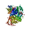
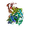
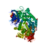
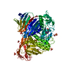
- PDB-5npc: Crystal Structure of D412N nucleophile mutant cjAgd31B (alpha-tra... -
+ Open data

- Basic information
| Entry | Database: PDB / ID: 5npc | ||||||
|---|---|---|---|---|---|---|---|
| Title | Crystal Structure of D412N nucleophile mutant cjAgd31B (alpha-transglucosylase from Glycoside Hydrolase Family 31) in complex with unreacted alpha Cyclophellitol Cyclosulfate probe ME647 | ||||||
 Components Components | Oligosaccharide 4-alpha-D-glucosyltransferase | ||||||
 Keywords Keywords | HYDROLASE | ||||||
| Function / homology |  Function and homology informationoligosaccharide 4-alpha-D-glucosyltransferase / oligosaccharide 4-alpha-D-glucosyltransferase activity / hydrolase activity, hydrolyzing O-glycosyl compounds / carbohydrate binding / carbohydrate metabolic process Function and homology informationoligosaccharide 4-alpha-D-glucosyltransferase / oligosaccharide 4-alpha-D-glucosyltransferase activity / hydrolase activity, hydrolyzing O-glycosyl compounds / carbohydrate binding / carbohydrate metabolic processSimilarity search - Function | ||||||
| Biological species |  Cellvibrio japonicus (bacteria) Cellvibrio japonicus (bacteria) | ||||||
| Method | X-RAY DIFFRACTION / SYNCHROTRON / MOLECULAR REPLACEMENT / Resolution: 1.96 Å | ||||||
 Authors Authors | Wu, L. / Davies, G.J. | ||||||
 Citation Citation | Journal: ACS Cent Sci / Year: 2017 Title: 1,6-Cyclophellitol Cyclosulfates: A New Class of Irreversible Glycosidase Inhibitor. Authors: Artola, M. / Wu, L. / Ferraz, M.J. / Kuo, C.L. / Raich, L. / Breen, I.Z. / Offen, W.A. / Codee, J.D.C. / van der Marel, G.A. / Rovira, C. / Aerts, J.M.F.G. / Davies, G.J. / Overkleeft, H.S. | ||||||
| History |
|
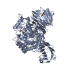
- Structure visualization
| Structure viewer | Molecule: MolmilJmol/JSmol |
|---|
- Downloads & links
-Download
| PDBx/mmCIF format | 5npc.cif.gz | 186.1 KB | Display | PDBx/mmCIF format |
|---|---|---|---|---|
| PDB format | pdb5npc.ent.gz | 142.8 KB | Display | PDB format |
| PDBx/mmJSON format | 5npc.json.gz | Tree view | PDBx/mmJSON format | |
| Others |  Other downloads Other downloads |
-Validation report
| Arichive directory | https://data.pdbj.org/pub/pdb/validation_reports/np/5npcftp://data.pdbj.org/pub/pdb/validation_reports/np/5npc | HTTPS FTP |
|---|
-Related structure data
| Related structure data | 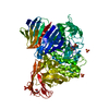 5npbC  5npdC 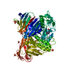 5npeC 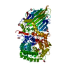 5npfC 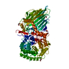 5o0sC  4b9yS C: citing same article ( S: Starting model for refinement |
|---|---|
| Similar structure data |


-Links
 PDBj
PDBj


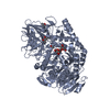
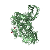
- Assembly
| Deposited unit | 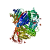
| ||||||||
|---|---|---|---|---|---|---|---|---|---|
| 1 |
| ||||||||
| Unit cell |
|
-Components
-Protein , 1 types, 1 molecules A
| #1: Protein | / Alpha-glucosidase 31B / CJAgd31B Mass: 94460.523 Da / Num. of mol.: 1 / Mutation: D412N Source method: isolated from a genetically manipulated source Source: (gene. exp.) Cellvibrio japonicus (bacteria) / Gene: agd31B, CJA_3248 / Production host: Escherichia coli (E. coli)References: UniProt: B3PEE6, oligosaccharide 4-alpha-D-glucosyltransferase |
|---|
-Non-polymers , 6 types, 385 molecules 


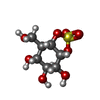
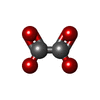

| #2: Chemical | ChemComp-SO4 / Sulfate Mass: 96.063 Da / Num. of mol.: 8 / Source method: obtained synthetically / Formula: SO4 Mass: 96.063 Da / Num. of mol.: 8 / Source method: obtained synthetically / Formula: SO4#3: Chemical | ChemComp-EDO / Ethylene glycol Mass: 62.068 Da / Num. of mol.: 12 / Source method: obtained synthetically / Formula: C2H6O2 Mass: 62.068 Da / Num. of mol.: 12 / Source method: obtained synthetically / Formula: C2H6O2#4: Chemical | ChemComp-PG4 / | Polyethylene glycol Mass: 194.226 Da / Num. of mol.: 1 / Source method: obtained synthetically / Formula: C8H18O5 / Comment: precipitant*YM Mass: 194.226 Da / Num. of mol.: 1 / Source method: obtained synthetically / Formula: C8H18O5 / Comment: precipitant*YM#5: Chemical | ChemComp-94E / ( |  Mass: 256.230 Da / Num. of mol.: 1 / Source method: obtained synthetically / Formula: C7H12O8S Mass: 256.230 Da / Num. of mol.: 1 / Source method: obtained synthetically / Formula: C7H12O8S#6: Chemical | ChemComp-OXL / | Oxalate Mass: 88.019 Da / Num. of mol.: 1 / Source method: obtained synthetically / Formula: C2O4 Mass: 88.019 Da / Num. of mol.: 1 / Source method: obtained synthetically / Formula: C2O4#7: Water | ChemComp-HOH / | WaterMass: 18.015 Da / Num. of mol.: 362 / Source method: isolated from a natural source / Formula: H2O |
|---|
-Experimental details
-Experiment
| Experiment | Method: X-RAY DIFFRACTION / Number of used crystals: 1 |
|---|
- Sample preparation
| Crystal | Density Matthews: 3.05 Å3/Da / Density % sol: 59.7 % |
|---|---|
| Crystal grow | Temperature: 293 K / Method: vapor diffusion Details: 1.8 M AMMONIUM SULFATE, 0.1 M HEPES (PH 7.0), 2% PEG400 |
-Data collection
| Diffraction | Mean temperature: 100 K |
|---|---|
| Diffraction source | Source: SYNCHROTRON / Site: Diamond  / Beamline: I02 / Wavelength: 0.97949 Å / Beamline: I02 / Wavelength: 0.97949 Å |
| Detector | Type: DECTRIS PILATUS 6M-F / Detector: PIXEL / Date: May 8, 2016 |
| Radiation | Protocol: SINGLE WAVELENGTH / Monochromatic (M) / Laue (L): M / Scattering type: x-ray |
| Radiation wavelength | Wavelength: 0.97949 Å / Relative weight: 1 |
| Reflection | Resolution: 1.96→65.67 Å / Num. obs: 84174 / % possible obs: 99.9 % / Redundancy: 20 % / CC1/2: 0.999 / Rmerge(I) obs: 0.2 / Rpim(I) all: 0.046 / Net I/σ(I): 11.4 |
| Reflection shell | Resolution: 1.96→2.01 Å / Redundancy: 19.2 % / Rmerge(I) obs: 2.991 / Mean I/σ(I) obs: 1.2 / Num. unique obs: 6171 / CC1/2: 0.625 / Rpim(I) all: 0.698 / % possible all: 99.9 |
- Processing
| Software |
| ||||||||||||||||||||||||||||||||||||||||||||||||||||||||||||||||||||||||||||||||||||||||||||||||||||||||||||||||||||||||||||||||||||||||||||||||||||||||||||||||||||||||||||||||||||||
|---|---|---|---|---|---|---|---|---|---|---|---|---|---|---|---|---|---|---|---|---|---|---|---|---|---|---|---|---|---|---|---|---|---|---|---|---|---|---|---|---|---|---|---|---|---|---|---|---|---|---|---|---|---|---|---|---|---|---|---|---|---|---|---|---|---|---|---|---|---|---|---|---|---|---|---|---|---|---|---|---|---|---|---|---|---|---|---|---|---|---|---|---|---|---|---|---|---|---|---|---|---|---|---|---|---|---|---|---|---|---|---|---|---|---|---|---|---|---|---|---|---|---|---|---|---|---|---|---|---|---|---|---|---|---|---|---|---|---|---|---|---|---|---|---|---|---|---|---|---|---|---|---|---|---|---|---|---|---|---|---|---|---|---|---|---|---|---|---|---|---|---|---|---|---|---|---|---|---|---|---|---|---|---|
| Refinement | Method to determine structure: MOLECULAR REPLACEMENT Starting model: 4b9y Resolution: 1.96→65.67 Å / Cor.coef. Fo:Fc: 0.969 / Cor.coef. Fo:Fc free: 0.955 / SU B: 5.825 / SU ML: 0.144 / Cross valid method: THROUGHOUT / ESU R: 0.135 / ESU R Free: 0.131 / Details: HYDROGENS HAVE BEEN ADDED IN THE RIDING POSITIONS
| ||||||||||||||||||||||||||||||||||||||||||||||||||||||||||||||||||||||||||||||||||||||||||||||||||||||||||||||||||||||||||||||||||||||||||||||||||||||||||||||||||||||||||||||||||||||
| Solvent computation | Ion probe radii: 0.8 Å / Shrinkage radii: 0.8 Å / VDW probe radii: 1.2 Å | ||||||||||||||||||||||||||||||||||||||||||||||||||||||||||||||||||||||||||||||||||||||||||||||||||||||||||||||||||||||||||||||||||||||||||||||||||||||||||||||||||||||||||||||||||||||
| Displacement parameters | Biso mean: 38.436 Å2
| ||||||||||||||||||||||||||||||||||||||||||||||||||||||||||||||||||||||||||||||||||||||||||||||||||||||||||||||||||||||||||||||||||||||||||||||||||||||||||||||||||||||||||||||||||||||
| Refinement step | Cycle: 1 / Resolution: 1.96→65.67 Å
| ||||||||||||||||||||||||||||||||||||||||||||||||||||||||||||||||||||||||||||||||||||||||||||||||||||||||||||||||||||||||||||||||||||||||||||||||||||||||||||||||||||||||||||||||||||||
| Refine LS restraints |
|