 Movie
Movie Controller

[English] 日本語
 Yorodumi
- PDB-9b37: Open state of kainate receptor GluK2 in complex with agonist glut... -
+ Open data

- Basic information
| Entry | Database: PDB / ID: 9b37 | |||||||||||||||
|---|---|---|---|---|---|---|---|---|---|---|---|---|---|---|---|---|
| Title | Open state of kainate receptor GluK2 in complex with agonist glutamate and positive allosteric modulator BPAM344 bound to one concanavalin A dimer. Composite map. | |||||||||||||||
 Components Components |
| |||||||||||||||
 Keywords Keywords |  MEMBRANE PROTEIN / kainate receptor / GluK2 / positive allosteric modulator / BPAM344 / open / concanavalin A / ConA / glutamate MEMBRANE PROTEIN / kainate receptor / GluK2 / positive allosteric modulator / BPAM344 / open / concanavalin A / ConA / glutamate | |||||||||||||||
| Function / homology |  Function and homology informationmossy fiber rosette / detection of cold stimulus involved in thermoception / Activation of Na-permeable kainate receptors / kainate selective glutamate receptor complex / Activation of Ca-permeable Kainate Receptor / negative regulation of synaptic transmission, glutamatergic / regulation of short-term neuronal synaptic plasticity / D-glucose binding / inhibitory postsynaptic potential / glutamate receptor activity ...mossy fiber rosette / detection of cold stimulus involved in thermoception / Activation of Na-permeable kainate receptors / kainate selective glutamate receptor complex / Activation of Ca-permeable Kainate Receptor / negative regulation of synaptic transmission, glutamatergic / regulation of short-term neuronal synaptic plasticity / D-glucose binding / inhibitory postsynaptic potential / glutamate receptor activity / ubiquitin conjugating enzyme binding / receptor clustering / modulation of excitatory postsynaptic potential / D-mannose binding / regulation of JNK cascade / kainate selective glutamate receptor activity / ionotropic glutamate receptor complex / extracellularly glutamate-gated ion channel activity / neuronal action potential / behavioral fear response / positive regulation of synaptic transmission / glutamate-gated receptor activity / ligand-gated monoatomic ion channel activity involved in regulation of presynaptic membrane potential / presynaptic modulation of chemical synaptic transmission / excitatory postsynaptic potential / dendrite cytoplasm / hippocampal mossy fiber to CA3 synapse / SNARE binding / synaptic transmission, glutamatergic / regulation of membrane potential / transmitter-gated monoatomic ion channel activity involved in regulation of postsynaptic membrane potential / PDZ domain binding / postsynaptic density membrane / regulation of long-term neuronal synaptic plasticity / modulation of chemical synaptic transmission / terminal bouton / intracellular calcium ion homeostasis / vasodilation / positive regulation of neuron apoptotic process / presynaptic membrane / manganese ion binding / scaffold protein binding / chemical synaptic transmission / postsynaptic membrane / perikaryon / neuron apoptotic process / negative regulation of neuron apoptotic process / postsynaptic density / axon / neuronal cell body / glutamatergic synapse / synapse / ubiquitin protein ligase binding / dendrite / calcium ion binding / membrane / identical protein binding / plasma membrane Function and homology informationmossy fiber rosette / detection of cold stimulus involved in thermoception / Activation of Na-permeable kainate receptors / kainate selective glutamate receptor complex / Activation of Ca-permeable Kainate Receptor / negative regulation of synaptic transmission, glutamatergic / regulation of short-term neuronal synaptic plasticity / D-glucose binding / inhibitory postsynaptic potential / glutamate receptor activity ...mossy fiber rosette / detection of cold stimulus involved in thermoception / Activation of Na-permeable kainate receptors / kainate selective glutamate receptor complex / Activation of Ca-permeable Kainate Receptor / negative regulation of synaptic transmission, glutamatergic / regulation of short-term neuronal synaptic plasticity / D-glucose binding / inhibitory postsynaptic potential / glutamate receptor activity / ubiquitin conjugating enzyme binding / receptor clustering / modulation of excitatory postsynaptic potential / D-mannose binding / regulation of JNK cascade / kainate selective glutamate receptor activity / ionotropic glutamate receptor complex / extracellularly glutamate-gated ion channel activity / neuronal action potential / behavioral fear response / positive regulation of synaptic transmission / glutamate-gated receptor activity / ligand-gated monoatomic ion channel activity involved in regulation of presynaptic membrane potential / presynaptic modulation of chemical synaptic transmission / excitatory postsynaptic potential / dendrite cytoplasm / hippocampal mossy fiber to CA3 synapse / SNARE binding / synaptic transmission, glutamatergic / regulation of membrane potential / transmitter-gated monoatomic ion channel activity involved in regulation of postsynaptic membrane potential / PDZ domain binding / postsynaptic density membrane / regulation of long-term neuronal synaptic plasticity / modulation of chemical synaptic transmission / terminal bouton / intracellular calcium ion homeostasis / vasodilation / positive regulation of neuron apoptotic process / presynaptic membrane / manganese ion binding / scaffold protein binding / chemical synaptic transmission / postsynaptic membrane / perikaryon / neuron apoptotic process / negative regulation of neuron apoptotic process / postsynaptic density / axon / neuronal cell body / glutamatergic synapse / synapse / ubiquitin protein ligase binding / dendrite / calcium ion binding / membrane / identical protein binding / plasma membraneSimilarity search - Function | |||||||||||||||
| Biological species |  Rattus norvegicus (Norway rat) Rattus norvegicus (Norway rat)  Canavalia ensiformis (jack bean) Canavalia ensiformis (jack bean) | |||||||||||||||
| Method | ELECTRON MICROSCOPY / single particle reconstruction / cryo EM / Resolution: 6.66 Å | |||||||||||||||
 Authors Authors | Nadezhdin, K.D. / Gangwar, S.P. / Sobolevsky, A.I. | |||||||||||||||
| Funding support |  United States, 4items United States, 4items
| |||||||||||||||
 Citation Citation | Journal: Nature / Year: 2024 Title: Kainate receptor channel opening and gating mechanism. Authors: Shanti Pal Gangwar / Maria V Yelshanskaya / Kirill D Nadezhdin / Laura Y Yen / Thomas P Newton / Muhammed Aktolun / Maria G Kurnikova / Alexander I Sobolevsky / Abstract: Kainate receptors, a subclass of ionotropic glutamate receptors, are tetrameric ligand-gated ion channels that mediate excitatory neurotransmission. Kainate receptors modulate neuronal circuits and ...Kainate receptors, a subclass of ionotropic glutamate receptors, are tetrameric ligand-gated ion channels that mediate excitatory neurotransmission. Kainate receptors modulate neuronal circuits and synaptic plasticity during the development and function of the central nervous system and are implicated in various neurological and psychiatric diseases, including epilepsy, depression, schizophrenia, anxiety and autism. Although structures of kainate receptor domains and subunit assemblies are available, the mechanism of kainate receptor gating remains poorly understood. Here we present cryo-electron microscopy structures of the kainate receptor GluK2 in the presence of the agonist glutamate and the positive allosteric modulators lectin concanavalin A and BPAM344. Concanavalin A and BPAM344 inhibit kainate receptor desensitization and prolong activation by acting as a spacer between the amino-terminal and ligand-binding domains and a stabilizer of the ligand-binding domain dimer interface, respectively. Channel opening involves the kinking of all four pore-forming M3 helices. Our structures reveal the molecular basis of kainate receptor gating, which could guide the development of drugs for treatment of neurological disorders. | |||||||||||||||
| History |
|
- Structure visualization
| Structure viewer | Molecule: MolmilJmol/JSmol |
|---|
- Downloads & links
-Download
| PDBx/mmCIF format | 9b37.cif.gz | 721.3 KB | Display | PDBx/mmCIF format |
|---|---|---|---|---|
| PDB format | pdb9b37.ent.gz | 604.3 KB | Display | PDB format |
| PDBx/mmJSON format | 9b37.json.gz | Tree view | PDBx/mmJSON format | |
| Others |  Other downloads Other downloads |
-Validation report
| Arichive directory | https://data.pdbj.org/pub/pdb/validation_reports/b3/9b37ftp://data.pdbj.org/pub/pdb/validation_reports/b3/9b37 | HTTPS FTP |
|---|
-Related structure data
| Related structure data |  44130MC 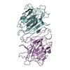 9b33C 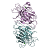 9b34C  9b35C  9b36C 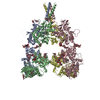 9b38C  9b39C C: citing same article ( M: map data used to model this data |
|---|---|
| Similar structure data |


-Links
 PDBj
PDBj


- Assembly
| Deposited unit | 
|
|---|---|
| 1 |
|
-Components
-Protein , 2 types, 6 molecules ABCDEF
| #1: Protein | Mass: 102976.586 Da / Num. of mol.: 4 Source method: isolated from a genetically manipulated source Source: (gene. exp.) Rattus norvegicus (Norway rat) / Gene: Grik2, Glur6 / Production host:  Homo sapiens (human) / References: UniProt: P42260 Homo sapiens (human) / References: UniProt: P42260#2: Protein | / ConVMass: 25622.385 Da / Num. of mol.: 2 / Source method: isolated from a natural source / Source: (natural) Canavalia ensiformis (jack bean) / References: UniProt: C0HJY1 |
|---|
-Sugars , 9 types, 29 molecules 
| #3: Polysaccharide | 2-acetamido-2-deoxy-beta-D-glucopyranose-(1-4)-2-acetamido-2-deoxy-beta-D-glucopyranose / Mass: 424.401 Da / Num. of mol.: 15Source method: isolated from a genetically manipulated source #4: Polysaccharide | / Mass: 586.542 Da / Num. of mol.: 3Source method: isolated from a genetically manipulated source #5: Polysaccharide | / Mass: 1479.349 Da / Num. of mol.: 2Source method: isolated from a genetically manipulated source #6: Polysaccharide | / Mass: 1276.157 Da / Num. of mol.: 2Source method: isolated from a genetically manipulated source #7: Polysaccharide | Cyclic 2-acetamido-2-deoxy-beta-D-glucopyranose-(1-2)-alpha-D-mannopyranose-(1-6)-beta-D- ...Cyclic 2-acetamido-2-deoxy-beta-D-glucopyranose-(1-2)-alpha-D-mannopyranose-(1-6)-beta-D-mannopyranose-(1-4)-2-acetamido-2-deoxy-beta-D-glucopyranose-(1-6)-[2-acetamido-2-deoxy-beta-D-glucopyranose-(1-2)]alpha-D-mannopyranose | Type: oligosaccharide / Mass: 1114.016 Da / Num. of mol.: 1Source method: isolated from a genetically manipulated source #8: Polysaccharide | / Mass: 748.682 Da / Num. of mol.: 2Source method: isolated from a genetically manipulated source #9: Polysaccharide | beta-D-mannopyranose-(1-3)-beta-D-mannopyranose-(1-4)-2-acetamido-2-deoxy-beta-D-glucopyranose-(1-4) ...beta-D-mannopyranose-(1-3)-beta-D-mannopyranose-(1-4)-2-acetamido-2-deoxy-beta-D-glucopyranose-(1-4)-2-acetamido-2-deoxy-beta-D-glucopyranose | / Mass: 748.682 Da / Num. of mol.: 1Source method: isolated from a genetically manipulated source #10: Polysaccharide | 2-acetamido-2-deoxy-beta-D-glucopyranose-(1-2)-alpha-D-mannopyranose-(1-3)-[2-acetamido-2-deoxy- ...2-acetamido-2-deoxy-beta-D-glucopyranose-(1-2)-alpha-D-mannopyranose-(1-3)-[2-acetamido-2-deoxy-beta-D-glucopyranose-(1-2)-alpha-D-mannopyranose-(1-6)]beta-D-mannopyranose-(1-4)-2-acetamido-2-deoxy-beta-D-glucopyranose-(1-4)-2-acetamido-2-deoxy-beta-D-glucopyranose | / Mass: 1317.209 Da / Num. of mol.: 1Source method: isolated from a genetically manipulated source #15: Sugar | N-Acetylglucosamine Type: D-saccharide, beta linking / Mass: 221.208 Da / Num. of mol.: 2 / Source method: obtained synthetically / Formula: C8H15NO6 Type: D-saccharide, beta linking / Mass: 221.208 Da / Num. of mol.: 2 / Source method: obtained synthetically / Formula: C8H15NO6 |
|---|
-Non-polymers , 6 types, 34 molecules 


| #11: Chemical | ChemComp-2J9 /  Mass: 242.270 Da / Num. of mol.: 4 / Source method: obtained synthetically / Formula: C10H11FN2O2S / Feature type: SUBJECT OF INVESTIGATION Mass: 242.270 Da / Num. of mol.: 4 / Source method: obtained synthetically / Formula: C10H11FN2O2S / Feature type: SUBJECT OF INVESTIGATION#12: Chemical | ChemComp-GLU / Glutamic acid Type: L-peptide linking / Mass: 147.129 Da / Num. of mol.: 4 / Source method: obtained synthetically / Formula: C5H9NO4 / Feature type: SUBJECT OF INVESTIGATION Type: L-peptide linking / Mass: 147.129 Da / Num. of mol.: 4 / Source method: obtained synthetically / Formula: C5H9NO4 / Feature type: SUBJECT OF INVESTIGATION#13: Chemical | ChemComp-POV / ( POPC Mass: 760.076 Da / Num. of mol.: 14 / Source method: obtained synthetically / Formula: C42H82NO8P / Comment: phospholipid*YM Mass: 760.076 Da / Num. of mol.: 14 / Source method: obtained synthetically / Formula: C42H82NO8P / Comment: phospholipid*YM#14: Chemical | ChemComp-CLR / Cholesterol Mass: 386.654 Da / Num. of mol.: 8 / Source method: obtained synthetically / Formula: C27H46O Mass: 386.654 Da / Num. of mol.: 8 / Source method: obtained synthetically / Formula: C27H46O#16: Chemical |  Mass: 65.409 Da / Num. of mol.: 2 / Source method: obtained synthetically / Formula: Zn Mass: 65.409 Da / Num. of mol.: 2 / Source method: obtained synthetically / Formula: Zn#17: Chemical |  Mass: 40.078 Da / Num. of mol.: 2 / Source method: obtained synthetically / Formula: Ca Mass: 40.078 Da / Num. of mol.: 2 / Source method: obtained synthetically / Formula: Ca |
|---|
-Details
| Has ligand of interest | Y |
|---|
-Experimental details
-Experiment
| Experiment | Method: ELECTRON MICROSCOPY |
|---|---|
| EM experiment | Aggregation state: PARTICLE / 3D reconstruction method: single particle reconstruction |
- Sample preparation
| Component | Name: full-length rat GluK2 tetramer in complex with one concanavalin A homodimer Type: COMPLEX / Entity ID: #1-#2 / Source: MULTIPLE SOURCES | ||||||||||||||||||||||||||||||||||||||||
|---|---|---|---|---|---|---|---|---|---|---|---|---|---|---|---|---|---|---|---|---|---|---|---|---|---|---|---|---|---|---|---|---|---|---|---|---|---|---|---|---|---|
| Molecular weight | Value: 0.46 MDa / Experimental value: NO | ||||||||||||||||||||||||||||||||||||||||
| Source (natural) | Organism: Rattus norvegicus (Norway rat) | ||||||||||||||||||||||||||||||||||||||||
| Source (recombinant) | Organism: Homo sapiens (human) / Cell: Human embryonic kidney 293 / Plasmid: pEG BacMam | ||||||||||||||||||||||||||||||||||||||||
| Buffer solution | pH: 8 | ||||||||||||||||||||||||||||||||||||||||
| Buffer component |
| ||||||||||||||||||||||||||||||||||||||||
| Specimen | Embedding applied: NO / Shadowing applied: NO / Staining applied: NO / Vitrification applied: YES | ||||||||||||||||||||||||||||||||||||||||
| Specimen support | Grid type: UltrAuFoil R1.2/1.3 | ||||||||||||||||||||||||||||||||||||||||
| Vitrification | Cryogen name: ETHANE |
- Electron microscopy imaging
| Experimental equipment |  Model: Titan Krios / Image courtesy: FEI Company |
|---|---|
| Microscopy | Model: TFS KRIOS |
| Electron gun | Electron source: FIELD EMISSION GUN / Accelerating voltage: 300 kV / Illumination mode: FLOOD BEAM |
| Electron lens | Mode: BRIGHT FIELDBright-field microscopy / Nominal defocus max: 2000 nm / Nominal defocus min: 1000 nm / Cs: 2.7 mm |
| Image recording | Average exposure time: 2.5 sec. / Electron dose: 58 e/Å2 / Film or detector model: GATAN K3 (6k x 4k) / Num. of grids imaged: 5 / Num. of real images: 22990 |
| Image scans | Width: 5760 / Height: 4092 |
- Processing
| EM software |
| ||||||||||||||||||||||||||||
|---|---|---|---|---|---|---|---|---|---|---|---|---|---|---|---|---|---|---|---|---|---|---|---|---|---|---|---|---|---|
| CTF correction | Type: NONE | ||||||||||||||||||||||||||||
| Particle selection | Num. of particles selected: 8660229 | ||||||||||||||||||||||||||||
| 3D reconstruction | Resolution: 6.66 Å / Resolution method: FSC 0.143 CUT-OFF / Num. of particles: 77567 Details: This is composite map. Reference map: 6.6 A ATD: 3.5 A LBD-TMD: 3.4 A ConA: 4.07 A Symmetry type: POINT | ||||||||||||||||||||||||||||
| Atomic model building | Space: REAL | ||||||||||||||||||||||||||||
| Refine LS restraints |
|
