 Movie
Movie Controller


+ Open data

- Basic information
| Entry | Database: PDB / ID: 8xck | |||||||||
|---|---|---|---|---|---|---|---|---|---|---|
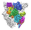
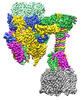
| Title | Closed state of central tail fiber of bacteriophage lambda | |||||||||
 Components Components |
| |||||||||
 Keywords Keywords |  VIRUS / Bacteriophage / caudovirales / siphoviridae / phage lambda / host recognition / LamB / cryo-EM VIRUS / Bacteriophage / caudovirales / siphoviridae / phage lambda / host recognition / LamB / cryo-EM | |||||||||
| Function / homology |  Function and homology information Function and homology informationsymbiont genome ejection through host cell envelope, long flexible tail mechanism / viral tail assembly / virus tail / peptidylprolyl isomerase / peptidyl-prolyl cis-trans isomerase activity / protein folding / outer membrane-bounded periplasmic space / host cell cytoplasm / entry receptor-mediated virion attachment to host cell / receptor-mediated virion attachment to host cell ...symbiont genome ejection through host cell envelope, long flexible tail mechanism / viral tail assembly / virus tail / peptidylprolyl isomerase / peptidyl-prolyl cis-trans isomerase activity / protein folding / outer membrane-bounded periplasmic space / host cell cytoplasm / entry receptor-mediated virion attachment to host cell / receptor-mediated virion attachment to host cell / periplasmic space / virion attachment to host cellSimilarity search - Function | |||||||||
| Biological species |  Escherichia phage Lambda (virus) Escherichia phage Lambda (virus) Escherichia coli (E. coli) Escherichia coli (E. coli) | |||||||||
| Method | ELECTRON MICROSCOPY / single particle reconstruction / cryo EM / Resolution: 2.75 Å | |||||||||
 Authors Authors | Ge, X.F. / Wang, J.W. | |||||||||
| Funding support |  China, 2items China, 2items
| |||||||||
 Citation Citation | Journal: To Be Published Title: Cryo-EM structure of lambda tail with LamB Authors: Ge, X.F. / Wang, J.W. | |||||||||
| History |
|
- Structure visualization
| Structure viewer | Molecule: MolmilJmol/JSmol |
|---|
- Downloads & links
-Download
| PDBx/mmCIF format | 8xck.cif.gz | 278.1 KB | Display | PDBx/mmCIF format |
|---|---|---|---|---|
| PDB format | pdb8xck.ent.gz | 220.4 KB | Display | PDB format |
| PDBx/mmJSON format | 8xck.json.gz | Tree view | PDBx/mmJSON format | |
| Others |  Other downloads Other downloads |
-Validation report
| Arichive directory | https://data.pdbj.org/pub/pdb/validation_reports/xc/8xckftp://data.pdbj.org/pub/pdb/validation_reports/xc/8xck | HTTPS FTP |
|---|
-Related structure data
| Related structure data | 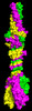 38246MC 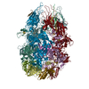 8xcgC 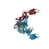 8xciC 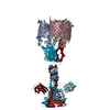 8xcjC M: map data used to model this data C: citing same article ( |
|---|---|
| Similar structure data |

-Links
 PDBj
PDBj

- Assembly
| Deposited unit | 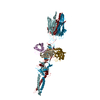
|
|---|---|
| 1 |
|
-Components
| #1: Protein | Mass: 46045.844 Da / Num. of mol.: 3 Source method: isolated from a genetically manipulated source Source: (gene. exp.) Escherichia phage Lambda (virus) / Production host: Escherichia coli (E. coli) / References: UniProt: P03749#2: Protein | Mass: 20453.277 Da / Num. of mol.: 3 Source method: isolated from a genetically manipulated source Source: (gene. exp.) Escherichia coli (E. coli) / Gene: ppiA, rot, rotA, b3363, JW3326 / Production host: Escherichia coli (E. coli) / References: UniProt: P0AFL3, peptidylprolyl isomerase |
|---|
-Experimental details
-Experiment
| Experiment | Method: ELECTRON MICROSCOPY |
|---|---|
| EM experiment | Aggregation state: PARTICLE / 3D reconstruction method: single particle reconstruction |
- Sample preparation
| Component |
| ||||||||||||||||||||||||
|---|---|---|---|---|---|---|---|---|---|---|---|---|---|---|---|---|---|---|---|---|---|---|---|---|---|
| Source (natural) |
| ||||||||||||||||||||||||
| Source (recombinant) | Organism: Escherichia coli (E. coli) | ||||||||||||||||||||||||
| Buffer solution | pH: 7 | ||||||||||||||||||||||||
| Specimen | Embedding applied: NO / Shadowing applied: NO / Staining applied: NO / Vitrification applied: YES | ||||||||||||||||||||||||
| Vitrification | Cryogen name: ETHANE |
- Electron microscopy imaging
| Experimental equipment |  Model: Titan Krios / Image courtesy: FEI Company |
|---|---|
| Microscopy | Model: FEI TITAN KRIOS |
| Electron gun | Electron source: FIELD EMISSION GUN / Accelerating voltage: 300 kV / Illumination mode: FLOOD BEAM |
| Electron lens | Mode: BRIGHT FIELDBright-field microscopy / Nominal defocus max: 2500 nm / Nominal defocus min: 1500 nm |
| Image recording | Electron dose: 50 e/Å2 / Film or detector model: GATAN K3 (6k x 4k) |
- Processing
| CTF correction | Type: NONE | ||||||||||||||||||||||||
|---|---|---|---|---|---|---|---|---|---|---|---|---|---|---|---|---|---|---|---|---|---|---|---|---|---|
| 3D reconstruction | Resolution: 2.75 Å / Resolution method: FSC 0.143 CUT-OFF / Num. of particles: 369942 / Symmetry type: POINT | ||||||||||||||||||||||||
| Refine LS restraints |
|
